
NEET Biology Muscular System
- Movement is the displacement of body parts in relation to the body axis. It is a characteristic feature of living beings. Movements of, external and internal body parts occur.
- Locomotion is the displacement of the body from one place to another place. Locomotion is the basic character of animals. Locomotion enables the animals to protect their bodies to search and procure food and water, to find their mate, escape from predators, shift to a favourable environment and find a suitable breeding ground.
- Locomotion in acellular organisms takes place by three methods :
- Pseudopodial
- Ciliary
- Flagellar.
- Pseudopodial locomotion takes place in Amoeba.
- Ciliary locomotion takes place in Paramecium.
- Flagellar locomotion takes place in Euglena.
- Adult sponges are sedentary ties. fixed. Locomotion does not take place in them.
- Their larvae move by cilia.
- Locomotion in Hydra is muscular and also takes place by tentacles,
- Bending and swaying
- Looping
- Somersaulting
Read and Learn More NEET Biology Notes
- Gliding
- Walking
- Climbing
- Surfacing
- Floating
- Swimming.
- Locomotion in annelids takes place either by setae or parapodia. Locomotion by setae takes place in Pheretima and by parapodia in Nereis.
- Locomotion in leeches takes place by muscular contraction and suckers.
- Locomotion in arthropods takes place by jointed legs and wings.
- Locomotion in molluscs takes place by foot
- Locomotion in echinoderms takes place by tube feet
- Locomotion in vertebrates takes place by fins and limbs.
NEET Biology Muscular System Notes
NEET Biology Kingdom Protista Movements of External Body Parts
- Collection of information by the movement of pinnae, ocular orbits, neck, etc.
- Facial expressions and gestures
- Preservation of balance
- Movement of extremities and limbs for locomotion.
- Acquisition of sustenance, Consumption of nourishment.
- Nourishing offspring, Offensive and Defensive
- Respiration.
NEET Biology Kingdom Protista Movements of Internal Body Parts
- The heartbeat facilitates blood circulation.
- Contraction and relaxation of the diaphragm, ribs, and lungs facilitate gaseous exchange.
- Peristalsis is the pendular or segmental motion facilitating the transit of food through the alimentary canal.
- Peristalsis facilitates the movement of urine through the urinary system, while also generating sound.
- Movements of the genital tract and uterine wall during oviposition and parturition.
- Muscular activity in the movements of different body components.
NEET Biology Kingdom Protista Non Muscular Movements
- Such movements occur in protists and individual cells in higher organisms in the following ways:
- Pseudopodial/Amoeboid Movements. The movements occur using temporary protoplasmic outgrowths called pseudopodia.
- Pseudopodia can develop in any direction due to the flow of protoplasm. Fresh pseudopodia develop as older ones are withdrawn.
- Amoeba and iLs relatives creep using pseudopodia. The movements occur in macrophages and leucocytes. Because of them the cells can reach all body parts and engulf pathogens.
- Cyclosis/Cytoplasmic or Protoplasmic Streaming. It occurs only inside eukaryotic cells. Cyclosis helps in the circulation of materials in the cells.
- Flagellar Movements. Flagella perform locomotion In euglenoids and other flagellate pivots, Chlamydomonas and other unicellular/colonial motile green algae sperm, etc. Flagellate choanocytes bring about the circulation of water in the canal system of sponges.
- Ciliary Movements. Paramecium and other ciliates swim using cilia.
- Undulation movements due to plasma membrane as in fibroblasts.
Muscular System NEET Study Material
NEET Biology Kingdom Protista Types Of Muscles
- Three types of muscles have been categorized based on the structure of their fibres.
- Details discussed in a chapter on (Animal tissues)
- Skeletal muscles (Striated muscles). These generate external movement associated with the skeleton, remain attached to bone through the tendon, under conscious control and hence also termed as voluntary muscle. In lower magnification, these have transverse stripes hence also called striped or striated muscles.
- Smooth Muscles (Visceral muscles)
- Unstriped; hence structurally different from striated and cardiac muscles.
- Muscle cell spindle-shaped (not cylindrical) uninucleated each formed of single myoblast.
- Actin filaments remain attached, to dense bodies in the cytoplasm.
- There is no myofibril, no sarcomere and no Z-line.
- Actin and Myosin filaments are not arranged in any pattern hence no striations.
- Involuntary muscle, under ANS control, is found in visceral organs (visceral muscle).
- Cardiac muscle. This is also striated muscle but under involuntary control, hence has a rhythmic pattern of contraction with varying frequency.
- This is an untiring muscle (no fatigue) and keeps on acting lifelong.
- Being striated it is structurally similar to skeletal muscle except for the presence of (0 Cross fibres, besides parallel fibres
- Intercalated discs – which represent the joints of two myoblasts.
- The contraction pattern is rhythmic and the rate of contraction is controlled by the autonomic nervous system (ANS).
- They do not get fatigued, and work ceaselessly throughout their lives.
- Generates impulse-like nerve (i.e. the myogenic heart of vertebrates).
NEET Biology Kingdom Protista Chemical Composition Of Muscle Fibres
- Water 75%
- Proteins 20% (Myosin, actin, tropomyosin troponin complex and enzymes)
- Proteins of Muscles
- Actin, the filament'(F-actin) is formed by polymerization of G-actin monomer. It has the site, occupied by regulatory protein troponin and tropomyosin, for the attachment of myosin.
- Tropomyosin is a rod-shaped fibrous protein helically wrapped around the actin filament along its groove.
- Troponin is a small globulin protein with l components: troponin-e, troponin-l and troponin-T masking the site on F-mMin lor attachment of myosin.
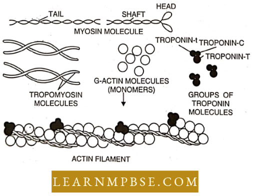
- Myosin is a versatile protein with both fibrous and globular parts. The fibrous part aggregates to find the filament while the globular part, projects out at both ends as cross bridges or heads. 1 lead is formed of HMM (heavy mcromysoin) consisting of Sub-fragment-1 and the neck (or hinge). The tail part is formed of LMM (light meromyosin). The head acts as ATPnse which after connecting with actin hydrolyse ATP.
- Mineral and organic compounds 5% (K+ is the chief mineral constituent, Mg++, P++, Na+ and Ca+ 4 are present in minute quantities.
- Organic compounds are present in the form of glycogen, lipids, steroids and non-poric nitrogenous compounds.
NEET Biology Kingdom Protista Some Major Muscles
- Biceps. The front portion of the upper ami for bending of the arm at the elbow.
- Triceps. Back of upper arm for straightening of ami at the elbow.
- Biceps femoris. Back of thigh for bending leg at the knee.
- Quadriceps. Front of thigh
- Gluteus maximus. Buttocks.
- Gastrocnemius. Calves, bending of the ankle joint.
- Frontal. Fore-head.
- Orbicularis oculi. Closure of eyes.
- Orbicularis oris. Closure of mouth.
- Masseter. Mastication.
- Trapezius. Upper back and side of the neck
- Deltoid. Shoulder raises an arm.
- Pectoralis major. Lowers arm.
NEET Biology Muscular System Notes
NEET Biology Kingdom Protista Features Of Muscle Fibers
- Excitability. The skeletal muscle fibres can be excited by the nerve impulses generated in the motor nerve fibre by the specific stimuli. This power is maximum in striated muscle fibres.
- Conductivity. The skeletal muscle fibres can conduct the excitation along the length of muscle fibres at the rate of 3-5 metres/second with the help of the T-tubule system and sarcoplasmic reticulum.
- All or None Rule (Bowditch’s Law). When the skeletal muscle fibre is stimulated, it responds either by shortening or lengthening. The response of a muscle fibre is independent of the strength of the stimulus. If there is any response, it is maximum.
- The minimum strength of stimulus (nerve impulse) required to bring about the response, is called threshold stimulus. It varies from fibre to fibre. If the stimulus is of a strength below the threshold stimulus, the muscle fibre is not excited, but if the stimulus has a strength equal to or higher than the threshold stimulus, the muscle fibre shows a maximum response.
- The force of contraction does not increase with the increase in the strength of the stimulus. But in the case of whole muscle, the extent of contraction depends upon the number of motor units which are contracting at any particular period.
NEET Biology Kingdom Protista Useful Terms
- Latent phase or period. It is a brief time interval between the point of stimulation and the actual starting of response i.e. contraction phase. The duration of the latent period varies with the species, type of muscle, temperature and internal condition of the muscle.
- It is due to the time taken for the propagation of impulse from the point of stimulation to the” neuromuscular junction and then to sarcolemma for initiation of contraction (In frogs it lasts about 10 milliseconds in skeletal muscles and up to 3 sec. in visceral muscles.)
- Contraction phase. It is the time interval in which the muscle reaches the peak of contraction and performs work. (It lasts for 40 milliseconds in skeletal and 20 sec. in visceral muscles.)
- Relaxation phase. It is the longest of three phases. It lasts about 50 milliseconds in skeletal muscles, and 2.3 sec in visceral muscles and is indicated by downward tracing in the kymograph (graph showing a twitch contraction). During the phase, the muscle returns to its original length.
- Single Muscle Twitch. The response of a muscle to a single short stimulus, such as 1 electric shock is known as a twitch and can be recorded as a kymograph. A twitch can be divided into three successive phases.
- Motor unit. All muscle fibres supplied by a single motor neuron are called a motor unit. On average, there are 100 muscle fibres in a motor unit.
- Isotonic contractions. When a muscle contracts, with a constant load that it can lift, it is said to be isotonic contraction In other words there is a change in the shape of the muscle and it maintains tension
- Isometric contractions. When a muscle contracts against a weight dial it cannot lift, it is called an isometric contraction. In other words, llicic is no change in the shape of the muscle. AS u maintains umfoim length.
- Tentas. It the muscle fibre is stimulated before it relaxes for a second time, it can contract again If we continue closely spaced stimuli, we gel a smooth sustained contraction, called a tetanus or tetanic contraction.
- Summation. If a second stimulus is applied to a muscle that is still in the contraction phase, a second contraction is added to die first and results in greater shortening of muscle than caused by a single stimulation. It is termed summation.
- Refractory period. (0.002 – 0.005 see in skeletal, 0.1-02 sec in cardiac muscle fibre.) If two stimuli are applied one immediately after the other, die muscle will respond to the first stimulus, but not to the second.
- The brief period during which the muscle does not respond to the second stimulus is called a refractory period or period of lost irritability. Its duration varies with the muscle involved. Skeletal muscle has a short refractory period w whereas cardiac muscle has a long refractory period.
- The absolute refractory period during which the recovering muscle fibre cannot be stimulated by a stimulus of any strength.
- The relative refractory period during which the muscle fibre can be excited only by a stimulus of strength higher than the previous strength of the stimulus.
- Fatigue. If muscle fibres are repeatedly stimulated to contract, these fibres take a longer time to respond to the excitation during contraction and also to complete relaxation.
- Their force of contraction also declines progressively. Finally, the fibres fail to contract at all for some time. It is known as muscle fatigue. It is caused by the accumulation of lactic acid and other changes in the muscle due to prolonged contraction.
- Site of muscle fatigue. Stephens and Taylor suggested that the site of skeletal muscle fatigue is the neuro-muscular junction and occurs when there is increased K+ concentration and decreased Na+ concentration in the ECF of the muscle fibres.
- Rigor mortis. Muscles become stiff after death. This state is called rigor mortis. It occurs due to the absence of ATP. It first appears in the muscles of the face and jaw. Rigour mortis disappears after three or four days because the proteins become soluble.
- Red muscle fibres contain myoglobin, arc thin, dark red and contain mitochondria. These are capable of doing more work and do not easily get tired.
- White muscle fibres are of light red or pink and do not contain myoglobin and mitochondria. Get easily tired and cannot work for long.
NEET Biology Kingdom Protista Muscle Contraction (Sliding Filament Theory)
- The sequence of myosin-actin bonding, sliding and release is repeated rapidly over and over until the muscle has shortened sufficiently. This is the sliding fuming mechanism for muscle contraction, and it consumes a great deal of ATP.
- When an individual muscle is stimulated, the actin filament in every sarcomere unit slides towards the middle of the sarcomere. As a result, the myofibrils shorten. A muscle cell shortens (contracts) then all its myofibrils shorten. An entire muscle contracts when many of its muscle cells contract simultaneously.
- Contraction ends when the muscle cell ceases to be stimulated by the nerve cell. If an impulse no longer travels down the T – tubules to the cell’s interior, Ca2+ is actively transported back into the SR.
- Without Ca2+, the troponin-tropomyosin protein complex shifts back into its original position, masking the sites on the actin filaments and preventing further bonding by the myosin heads. When this happens, the muscle relaxes and returns to its original shape.
NEET Biology Kingdom Protista Biochemical Aspects
- During contraction actin and myosin combine to form actomyosin, this was shown in vitro by Szent Gyorgi (1946), who won the Nobel Prize for this work.
- There are three basic pathways for providing energy to be utilised in muscle contraction.
- The hydrolysis of ATP provides free energy to the converted to mechanical energy by the interaction of actin and myosin. Here myosin plays the role of ATPase.
- ADP + CP + H+ → ADP + Pi + H+ ΔG
- The ATP molecule present in muscle cells lasts for only a few seconds of muscle action. Hence muscle mainly depends upon other phosphate molecules creatine phosphate (CP) as a source of energy stored it. This acts as a recharger of ADP. ADP + CP + H+ →ATP + C (Lohmann’s Reaction)
- This reaction was discovered by Lohmann (1932) which won him a Nobel Prize.
- After the CP of reserve is exhausted muscle resorts to glycolysis of glycogen. Whose end product is the lactic acid that causes fatigue.
- Oxygen debt. Till the entire amount of creatine phosphate is recovered through the regeneration of ATP by oxidative phosphorylation body remains in oxygen debt and to repay it the person keeps on panting after a strenuous exercise. Cori cycle. The accumulated lactic acid diffuses out of muscle through blood and reaches the liver where it is reconverted to glycogen.
- Troponin and Tropomyosin are two regulatory proteins present on this filament and are essential for the actin-myosin interaction i.e., muscle contraction.
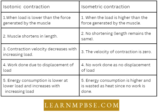

- When during muscle contraction, muscle length remains c& the same but tension increases then it is called isometric contraction For Example Holding some weight.
- When during muscle contraction, muscle length changes but tension remains constant then it is called isotonic contraction For Example during walking. c& Glycogen can be formed more readily from glucose in muscles.
- Carl Ferdinand Cori shared the 1947 Nobel Prize for physiology and medicine with his wife Gerty Theresa Cori and B.A. Houssary for their work on the metabolism of glycogen.
- Animals with“liquid skeleton”-Earthworms, Caterpillars (larvae of butterflies and silk moths), and Slugs. It gives shape and support to the body. c& In the case of non-chordates except echinoderms arginine is present in place of phosphocreatine.
- Sarcomere functional unit of contraction of a muscle.
- All or none law is also known as Bowditch’s law. Biochemical and electrical events during muscle contraction were studied by Albert Szent Gyorgi Special conducting system of the heart consists of special cardiac muscle fibres.
- Treppe (Staircase phenomenon) A stimulus of constant strength applied regularly at clock intervals to a muscle produces a series of contractions, the first of which are of increasing amplitude.
- Glycogenolysis occurs in liver cells but not in muscle cells as these lack the Glucose-6 phosphatase enzyme. Troponin-Tropomyosin Complex prevents muscle contraction as it inhibits the activity of ATPase enzyme and prevents the formation of actomyosin complexes., Oxygen-Debt. Amount of extra oxygen consumed during the recovery after an exercise.
- Shivering involves the involuntary contraction of voluntary muscle fibres.
- 30-40 % of energy is used in contraction and the rest is converted into heat. This helps in warming the body.
- Roles of Muscle and Bones in Movement The force generated by muscle contraction pulls or lifts bone through tendons. Movement occurs along the joint, hence the bone actually acts as a lever having 3 parts :
- In the body joint acts as F, point of attachment of muscle to bone is E.
- There are 3 classes of levers:
- 1st class lever. F comes between E and R, very rare in the body, one such example is the head resting on the vertebral column.
- 2nd class lever. R comes between F and E, only a few in the body, For Example, raising the body on the toes.
- 3rd class lever. E remains in between the F and R, the most common type of lever in the body For Example, flexing the forearm at the elbow joint.
NEET Biology Muscular System Notes
NEET Biology Kingdom Protista Questions From Competitive Examinations
Question 1. Immediate Source Of Energy For Muscle Contraction Is:
- Glucose
- Gtp
- Creatine Phosphate
- ATP
Answer: 4. ATP
Question 2. Which One Yields ATP Required For Muscle Contraction?
- Myoglobin
- Creatine Phosphate
- Creatinine Phosphate
- Myosin.
Answer: 2. Creatine Phosphate
Question 3. Edta Injected Into Muscles Combines With Ca2+ And :
- Stops Contraction
- Causes Contraction
- Slows Down Contraction
- None Of The Above.
Answer: 1. Stops Contraction
Question 4. What Is Sarcomere?
- Part Between Two H-Line
- Part Between Two A-Line
- Part Between Two I-Band
- Part Between Two Z-Iine.
Answer: 4. Part Between Two Z-Iine.
Question 5. Which Statement Is Correct For Muscle Contraction?
- Length Of H-Zone Becomes Decreased
- Length Of A-Band Remains Constant
- Length Of I-Band Becomes Increased
- Length Of Two Z-Line Become Increase,
Answer: 1. Length Of H-Zone Becomes Decreased
Types of Muscles NEET
Question 6. Which light band Contains The following fluid protein?
- Myosin
- Actin
- Both 1 & 2
- None Of These.
Answer: 2. Actin
Question 7. Largest Skeletal Muscle Is Present In :
- Thigh
- Neck
- Arm
- Belly.
Answer: 1. Thigh
Question 8. Hinge Joint Is Present In :
- Elbow
- Pivot
- Interphalangeal
- Atlas.
Answer: 1. Elbow
Question 9. Biceps & Triceps Are Found In :
- Fore Arm
- Upper Arm
- Thigh
- Shoulder.
Answer: 2. Upper Arm
Question 10. Actin Filaments Depolymerise And Repolymerise In Motile Cells With :
- 0.2 – 0.3 Millisecond
- 1.0 – 2.0 Minutes
- 1 – 2 Hours
- 2-3 Days.
Answer: 1. 0.2 – 0.3 Millisecond
Question 11. Sacromere Occurs Between :
- Z – Line To Z – Line
- Two I – Bands
- Two Surfaces Of A Z – Line
- Two A – Bands.
Answer: 1. Z – Line To Z – Line
Question 12. “All Or None Law” Is Not Associated With.
- Muscle Fibre
- Tiruiun
- Cardiac Muscle
- Uriniferous Tubules
Answer: 4. Uriniferous Tubules
Question 13. ATPase Enzyme Needed For Muscle Contraction Is Located In :
- Myosin
- Actin
- Actinin
- Troponin.
Answer: 1. Myosin
Types of Muscles NEET
Question 14. Which Of The Following Is Important For Muscle Contraction And Nerve Impulse Transmission?
- Ca2+ Ions
- Ca++ And Mg++ Ions
- Mg+ 4– Ions
- Fe 2+ Ions.
Answer: 2. Ca++ And Mg++ Ions
Question 15. During Strenuous Exercise Glucose Is Converted Into :
- Glycogen
- Pyruvic Acid
- Starch
- Lactic Acid.
Answer: 4. Lactic Acid.
Skeletal Muscle Structure NEET
Question 16. Which Of The Following Is Not Syncytial?
- Cardiac Muscle
- Skeletal Muscle
- Smooth Muscle
- Interstitial Muscle.
Answer: 3. Smooth Muscle
Question 17. The Condition In Which A Number Of Nuclei Are Present In A Muscle Fibre Is Called :
- Coenocytic
- Syncytial
- Polykaryon
- Endoduplicaton.
Answer: 2. Syncytial
Question 18. Contractile Tissues Have The Following Features :
A. Mesodermal In Orign
B. They Contain Stretch Receptors.
C. Rhythmic Contractions Are Seen In Them
D. Hey Do Not Fatigue During The Life Of The Animal.
Which Of The Above Are Characteristics Of Sphincters?
- All The Four
- Only A, B And C
- Only A, B And D
- Only A, C And D.
Answer: 2. Only A, B And C
Question 19. The Lactic Acid Generated During Muscle Contraction Is Converted To Glycogen In :
- Muscle
- Kidney
- Pancreas
- Liver.
Answer: 2. cAMP
Question 20. In Heart Cells, Which One Serves As A Second Messenger, Speeding Up Muscle Cell Contraction In Response To Adrenaline?
- cAMP
- cGMP
- GTP
- ATP
- AMP.
Answer: 1. cAMP
Skeletal Muscle Structure NEET
Question 21. The Contractile Protein Of Skeletal Muscle Involving Atpase Activity Is :
- Troponin
- A-Actin
- Tropomyosin
- Myosin.
Answer: 4. Myosin.
Question 22. The Globular Protein Molecule, Which Masks The Active Sites On The F-Actin Is :
- Troponin
- Myosin
- Tropomyosin
- Light Mbromyosin
- Heavy Meromyosin.
Answer: 1. Troponin
Question 23. Statements :
- A-Bands Of The Muscle Is Dark And Contain Myosin
- I-Hamls Air The Light Hands Ami Contain Actin
- During Muscle Contraction The A-band Contracts
- The Part Between The Two Z-Lines Is Called As Sacromere
- The Central Pan Of Thin Filament, Not Overlapped By Thick Filament Is Called H-Zone.
Of The Above Statements.
- 1, 3, and 5 Are Correct While 2 And 4 Are Incorrect
- 1, 2, 3 And 3 Are Correct While 4 Is Incorrect
- 1, 2 And 3 Are Correct While 4 And 3 Are Incorrect
- 1, 2 And 4 Are Correct While 3 And 3 Are Incorrect
- 1 And 2 Are Correct While 3, 4 And 5 Are Incorrect.
Answer: 4. 1, 2 And 4 Are Correct While 3 And 3 Are Incorrect
Question 24. All Or None Law Is Not Applicable To :
- Whole Skeletal Muscle
- Single Skeletal Muscle Fibre
- Whole Cardiac Muscle
- Single Smooth Muscle Fibre.
Answer: 1. Whole Skeletal Muscle
Question 25. The H-Zonc In The Skeletal Muscle Fibre Is Due To :
- The Central Gap Between Myosin Filaments In The A-Band.
- The Central Gap Between Actin Filaments Extending Through Myosin Filaments In The A Band.
- Extension Of Myosin Filaments In The Central Portion Of The A-Band.
- The Absence Of Myofibrils In The Central Portion Of A-Band.
Answer: 2. The Central Gap Between Actin Filaments Extending Through Myosin Filaments In The A Band.