
Biomembranes And Cell Wall
The plasma membrane and intracellular membranes collectively constitute biological membranes.
- The plasma membrane surrounds each cell, defining its limits and maintaining essential separations between its internal components and the external environment. It exhibits selective permeability.
- Intracellular membranes partition the cell into functionally distinct compartments known as organelles, including mitochondria and chloroplasts.
- Biological membranes demonstrate selective permeability, permitting the passage of some molecules while inhibiting others.
- All biological membranes display dynamism.
- All biological membranes of eukaryotic cells have fundamental architecture, comprising lipids and protein molecules interconnected by non-covalent interactions.
- Chemical composition. The plasma membrane consists of 58% proteins, encompassing structural and functional proteins like permeases, and 40% lipids, mostly phospholipids, cholesterol, and galactolipids.
- Minor polymers of monosaccharides (L-27%) form weak interactions with proteins and lipids.
Function and Structure of Cell Wall Membrane For NEET Models Of The Structure Of Plasma Membrane
The plasma membrane under an electron microscope appears to be trilaminar. Davson and Danielli put forward the Lamellar model which states that it is formed of four layers i.e. two proteins and two phospholipids. J.D. Robertson gave the concept of unit membrane which means that all membranes have three layers with a phospholipid bilayer sandwiched between two layers of proteins.
Read and Learn More NEET Biology Notes
According to the unit-membrane concept, all cell membranes possess a common basic structure, consisting of two layers of proteins and a middle bilayer of lipids. According to the fluid-mosaic model, the lipids and proteins are managed in a mosaic fashion in the membrane.
The cell membrane possesses a quasi-fluid phospholipid layer with embedded proteins. These impart considerable fluidity to the membranes.
Singer and Nicholson proposed a Fluid mosaic model. According to this model, phospholipids are arranged in a bimolecular layer with their hydrophilic polar heads forming outer surfaces and hydrophobic non-polar tails buried in the membrane.
The proteins are extrinsic and intrinsic. The plasma membrane is in the form of quasi-fluid. It also explains the permeability of carrier proteins.
Cell Wall And Cell Membrane Neet Notes
Specialization of the plasma membrane. Various modifications are formed to perform special functions. They are microvilli. The adjacent cells may develop specialized structures such as interdigitations, desmosomes, intermediary junctions, and tight junctions.
Functions. lt provides shape and protects the contents inside the cell.
It controls permeation and has the property of retentivity. It binds the semifluid cellular constituents. It also acts as a barrier between protoplasm and the external environment. It possesses properties of permeability, and surface tension and is electrically charged.
Function and Structure of Cell Wall Membrane For NEET Flow Of Materials
To perform the various life activities, the cell must exchange certain substances with the environment.
It is necessary that metabolic essentials such as oxygen mineral salts and nutrients must get into the cell and waste products such as ammonia, COr, calcium crystals, and other metabolites, must pass out of the cell.
All these exchanges can take place only through the cell membrane by many processes as follows:
- Passive transport
- Diffusion
- Osmosis
- Facilitated diffusion
- Active transport
- Na+ and K+ exchange Pump
- Endocytosis
- Phagocytosis
- Pinocytosis
- Exocytosis
Transport of materials takes place across the membranes by two major processes-
Passive Transport and Active Transport. Passive transport is a physical process that neither requires energy nor oxygen for the transport of substances by the cell. This includes diffusion and osmosis’
Diffusion. The physical process involves the transport of materials from a region of higher concentration to a region (solutes and gases) of lower concentration and pressure respectively to spread uniformly.
Facilitated diffusion. A substance first combines with the carrier protein without the use of energy in the cell membrane and then it diffuses across the membrane.
The diffusion rate can be expressed by the formula:
Diffusion rate = Concentration difference x Cross-sectional area x Temperature/\tlolecular
weight x Distance x Density of Medium.
Osmosis. Bulk flow of water through a semipermeable membrane into another aqueous phase containing a solute in higher concentration Osmotic pressure.
Pressure is generated by the osmotic flow of water through a membrane into an aqueous phase containing a solute in higher concentration.
Structure And Function Of Cell Membrane Neet
Plasmolysis. A plant cell when placed in hypertonic solution undergoes the shrinkage of its cytoplasm with plasma membrane.
Plants can absorb water through osmosis Active transport is an energy-dependent process.
A membrane protein acts as a carrier that binds and transports the molecule or ion in a specific direction across the membrane It even takes place against an electrochemical gradient.
In sodium-potassium exchange pump Na+ – K+ ATPase actively transports three molecules of Na+ outwards and two molecules of K+ inward.
Endocytosis. Intaking of large-sized liquid or solid macromolecules inside the cell through the plasma membrane.
Exocytosis. (cell vomiting). Expelling of cellular secretions of gland cells or wastes out of the cell through the plasma membrane.
Phagocytosis (cell-eating). Ingestion of solid particles by the cell Phagocytosis is of two kinds viz.
Colloidopexy: In this case plasma membrane ingests small colloidal particles as shown by leucocytes and macrophage cells of mammals.
Chromopexy Colloidal chromogen particles are ingested by plasma membrane as shown by mesoblastic cells of mammals.
Pinocytosis is the process of endocytosis in which liquid food is engulfed in bulk through a plasma membrane.
Plant cells possess cell walls and this feature distinguishes them from animal cells.
The cell wall is partially non-living and partially living structure. It is secreted by cytoplasm or protoplast. It has a supportive and protective function.
There are three distinct layers -middle lamella, primary cell wall, and secondary cell wall. Occa- a tertiary cell wall may be present.
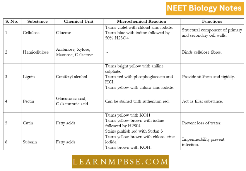
‘u’ Middle lamella is a cementing layer between the primary walls of adjacent cells. It is composed of pectin, calcium, and magnesium. During the ripening of fruits enzyme pectinase dissolves it.
The primary cell wall is 0.1pm thick. It is composed of chiefly cellulose, hemicellulose, and pectic compounds. Cellulose fibrils are made up of 8,000 to 12,000 glucose units, they are about 1004 in diameter.
The secondary wall is laid down inner to the primary wall. It is 5 to 10 pm thick.
The secondary cell wall has three layers- outer layer, middle layer, and inner layer. The primary wall has small pores called plasmodesmata which connect the cytoplasm of adjacent cells. The cell wall is made up of matrix and microfibrils.
Molecular organization. The cell wall appears to consist of a gelatinous amorphous matrix consisting of polysaccharides (mainly pectin), gums, tannins, silica, and wax.
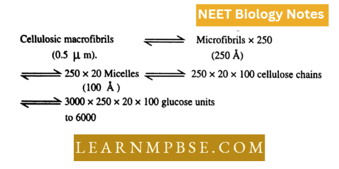
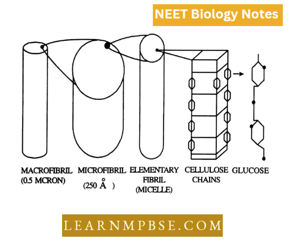
In the matrix are found many fibrils called microfibrils. 0.5 pm in width visible under a light microscope. The microfibril consists of bundles of 250 each, which is about 250 i in diameter. It is visible under an electron microscope.
The microfibril in turn consists of bundles of micelles elementary fibrils each about 100 i in diameter. The micelle contains about 100 cellulose chains.
Each microfibril is lpm long and 100-250 i in diuter. Each microfibrilconsists of 20 micelles. Each micelle consists of 100 individual cellulose chains. Each cellulose chain consists of 3000 – 6000 glucose molecules linked by B 1-4 linkage and is unbranched.
Origin. The cell wall is laid during the telophase stage of cell division from vesicles of the Golgi complex or E.R.
Function. The cell wall is a highly active region as many enzymatic activities are known to occur. In addition to providing shape and strength, it protects the cell organelles and plays a vital role in cell expansion. It helps in the transport of materials and metabolites in and out of the cell and encounters the turgor pressure developed inside.
- Most of the prokaryotic cells particularly bacterial cells, have a chemically complex cell envelope. Its layers are stacked upon one another.
- The outermost layer is glycocalyx followed by cell wall and cell membrane (plasma membrane).
- Glycocalyx. Any coat present outside the plasma membrane is called a cell coat. It forms a cell wall in plant cells and a pellicle in prokaryotes. In animal cells polysaccha- rides form the external cell coat or glycocalyx. This layer of glycocalyx differs in thickness and chemical composition in different bacteria. Some have a loose sheath called a slime layer which protects the cells from loss of nutrients and water. Some others have a tough covering called the capsule.
- In bacterial cells, the cell coat is formed of a protein-lipid polysaccharide complex.
- Bacteria can be classified according to their ability to take up and retain certain dyes.
- Gram staining is the most widely used staining procedure.
- The gram staining technique divides most bacteria into one or two groups.
- A Danish physician, Dr. Hans Christian Gram developed this staining method in 1884 to distinguish between the bacteria that cause pneumonia and eukaryotic cell nuclei in infected mammalian tissues.
- Differences between Gram-positive and Gram-negative Bacteria
- In Mycobacterium and Noccardia, the wall is of Gram-positive type but a part of their cell wall is made up of a very long chain of fatty acid called mycoic acid.
- Differences between pilus and fimbriae The Primary cell wall in plant cells is composed of an intricate network of microfibrils in a gel-like matrix arranged in cellulose microfibrils, pectic polysaccharides, and structural proteins.
- A new class of protein called expansion is responsible for wall loosening and cell expansion by the addition of cellulose molecules to cellulose microfibrils.
- The matrix of the wall contains water, pectin, hemicellulose, and glycoproteins. Pectin is the filler substance of the matrix. It is a mixture of polymerized and methylated galacturans, galacturonic acid, and neutral sugars.
- Glycoproteins control the orientation of microfibrils. They also exert enzymatic influence on the metabolic activities of the wall. Hemicellulose is a mixture of polymerized xylans, mannans, glucomannans, galactans, and arabinogalactans. It binds microfibrils with the matrix.
- The phospholipids and integral proteins of the plasma membrane are amphipathic. The term was coined by Hartley in 1936 for those molecules which have both hydrophobic and hydrophilic groups.
- The flexibility of the cell membrane is due to the presence of fatty acid contents. This enables bacteria to maintain the same fluidity over a temperature range of almost 100. c.
- The proteins are not easily extricated from the membrane because of their high degree of insolubility and non-covalent bonds.
- The plasmodesmata help in maintaining the continuity of living matter and cytoplasm, such condition is called sym-plasm. In contrast to this, the intercellular space containing non-living matter is called apoplasm.
Difference Between Cell Wall And Cell Membrane Neet
Quanta To Memory
- Membrane-less cell organelles include ribosomes, centrioles, centrosomes, nucleoli, microtubules, microfilaments, chromosomes, and intermediate filaments.
- Cell organelles characterized by a single membrane: lysosomes, Golgi apparatus, endoplasmic reticulum, thylakoids, peroxisomes, protoplasts, vacuoles, glyoxisomes, flagella, and cilia.
- Cell organelles have a double membrane: plastids, mitochondria, nucleus.
- Cell organelles possessing many membrane layers exceeding two.
- Tranosome — three membrane encasement.
- The plastids of Euglenoids and Dinoflagellates possess three membrane layers.
- Diatom plastids possess four membrane layers.
- The typical thickness of the plasma membrane is from 70 to 100 Å, but the plasma membrane of red blood cells measures 215 Å.
- The dimensions of molecules capable of traversing the plasma membrane range from 1 to 15 angstroms.
- The nuclear membrane creates outpockets that are constricted to produce blebs or vesicles, a phenomenon known as blebbing, as reported by Gay in Drosophila.
- The most intricate glycolipids are gangliosides, which incorporate one or more sialic acid residues, also referred to as N-acetyl neuraminic acid (NANA). It imparts a negative charge to the cell membrane.
- Glycoproteins facilitate cellular recognition. A membrane is predominantly maintained by hydrophobic interactions.
- The glycocalyx is an extracellular coating located outside the cell membrane in animal cells. It comprises oligosaccharides linked to lipids and proteins, resulting in glycolipids and glycoproteins. It serves a protective function and operates as a recognition hub.
- Lipid-soluble compounds traverse the plasma membrane more easily than water-soluble molecules.
- Protein-free lipid bilayers exhibit impermeability to ions while allowing unrestricted permeability to water.
- The secretion of neurotransmitters by neurons exemplifies exocytosis.
- Seagulls and penguins excrete NaCl via nasal glands containing Na+ – K+ pumps.
- Younger cells possess thinner and elastic (semi-rigid) cell walls to facilitate growth by intussusception.
- Meristematic, parenchymatous cells, and root hairs possess solely primary cell walls
Matrix is an amorphous, gel-like material. It has watery hemicellulose and pectin but also has lipids and protein. Hemicellulose is made of:
- Arabinose
- Mannose
- Pectin consists of glucuronic acid and galacturonic acid units.
- The tertiary wall is made of cellulose and xylan.
- Plasmodesmata are inner cytoplasmic strands.
- Ca++ cross-links play a role in holding cell wall components together.
- The cell wall is permeable as spaces of framework allow free passage of dissolved materials between cells and their environment. The cell coat is different from the cell wall.
- Water moves passively through the apoplast which gives thermo- charge.
- Messenger recognition sites of two neighboring cells may bind to each other causing cell adhesion.
- Glycophorin is present in the membrane of RBC.
- In Immune responses of various response systems, glycoproteins act as antigens.
- Lipid molecules have kinks in fatty acid tails which prevent the close packing of molecules and make the membrane structure more fluid. Fluidity increases, decreasing the length of fatty acid tails. Kinks are due to double bonds in the tail.
- Carbohydrates are attached to the polar side of the lipid to form Glyeolipid 1 he lipid: Protein ratio in some eases aw as under
- 40: 60
- 35: 65
- 42: 58
- 76: 24
Functions of proteins in Plasma membrane
- Transporters of molecules across cellular membranes.
- Receptors. Information transfer within cells.
- Enzymes. Each membrane contains enzyme molecules designated for specific cellular functions.
- The plasma membrane does not permit the escape of the cell’s contents, as it retains its color due to the presence of pigment molecules.
- Gases have the highest rate of diffusion. Ions and diminutive polar molecules disperse at a sluggish rate. Fat-soluble molecules have the highest rate of passage.
- Daniel and Davson’s model was proposed prior to the observation of the plasma membrane using an electron microscope.
- The fluid mosaic concept posits that the bilayer of phospholipid molecules acts as a relatively impermeable barrier to the transit of most water-soluble substances.
- It is proposed that water exists within the plasma membrane. In the absence of water, how do lipids develop hydrophilic and hydrophobic regions? Only recommended
- Plasma membrane organelles perform analogous activities due to their fundamental structural similarities. Nonetheless, many membranes exhibit numerous structural and physicochemical functional disparities.
- The ratio of lipids to proteins ranges from 1:08 to 1:4. The unit membrane idea cannot be applied unconditionally.
- The lignified wall is colored with safranin. The cell wall is lacking in mycoplasma, gametes, and animal cells.