NEET Biology Cell Unit Of Lift And Structural Organisation
All living organisms consist of many coordinated compartments known as cells.
- Robert Hooke (1665) was the inaugural individual to employ the term ‘cell.’
- In 1831, Robert Brown identified a nucleus within orchid roots.
- Cytology: The discipline focused on the scientific examination of cells is known as cytology.
- The inception occurred in the early 17th century with the initial utilization of microscopes by Hooke, Grew, and Malpighi.
- Cellular biology: A comprehensive examination of a cell, considering its structure, functions, biochemical characteristics, reproduction, energy dynamics, division, development, and differentiation.
- Cell theory: German scientists Schleiden and Schwann proposed the cell theory, which asserts that all living organisms consist of cells.
NEET Biology Cell Unit Of Life Present Day Concept Of Cell Theory
- The body of almost all living organisms is an assemblage of cells or the cell constitutes the structural unit of all living organisms.
- Each cell represents a unit of metabolic activities because all the characteristics of life are performed within the cell.
- Cells are hereditary units also as they contain hereditary materials inside the nucleus.
- New cells originate from pre-existing cells. (R. Virchow 1858).
- All the activities are the outcome of the activities of its constituent cells.
- All the cells are capable of independent existence and they work as autonomous units.
- In multicellular organisms, a clear-cut division of labour can be noticed. However, cells are still dependent partly on each other.
Read and Learn More NEET Biology Notes
Cell The Unit Of Life NEET Notes
Cellular totipotency is the ability of a somatic cell of a plant to produce a new complete plant.
- Steward et al. showed the phenomenon of cellular totipotency in carrot cultures,
- Unicellular organisms are capable of performing all the metabolic activities.
- For an efficient cell, the ratio of volume to surface area should be high.
- A cell represents itself in two forms i.e. as an individual and as part of the continuity of cells.
- Multicellular organisms are adapted for better survival.
- Growth and synthesis of protoplasm are due to the nucleus of the cell.
- Cytoplasm (mesoplasm cytosol or kinoplasm) is protoplasm except nucleus.
- Protoplasm is a reversible crystallocolloidal solution. Term cytoplasm was given by Kolliker.
- Hyaloplasm is cytoplasm outside the nucleus.
- Cytoplasm coagulate when treated with acids/bases or heated above 60°C.
- AH, physical and chemical changes tend to proceed in such a direction that useful energy undergoes irreversible degradation into a random form called entropy.
- pH (powered hydrogen) or puissance hydrogen of cytoplasm, nucleoplasm and human blood is 6.9 ± 0.2, 7.4 ± 0.2 and 7.34 ± 0.2 respectively.
NEET Biology Cell Unit Of Life Types Of Cell
Prokaryotic cell. A primitive cell which lacks a nuclear membrane, or nucleolus, possesses 70S ribosomes, membrane-bound organelles absent, and naked DNA present, also called a nucleoid or genophore or chromosome or fibrous nucleus or incipient nucleus, Example Bacteria, Blue green algae, PPLO etc.
Mesokaryotic cell. It is characterized by the absence of deoxyribounyleohistone; however, well well-defined nucleus is present. A peculiar type of division called dino mitosis occurs. Dodge used the term for cells of dinoflagellates (a type of algae). Eukaryotic cell.
Present in higher organisms. It possesses a true nucleus, DNA is associated with histone proteins called chromatin. Membrane-bound cell organelles present.

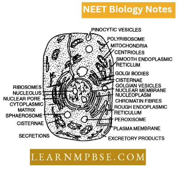
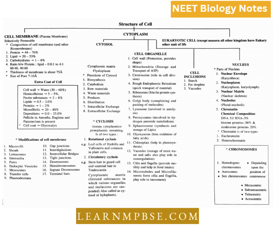
Cell Structure and Its organelles
- A cell is a specific combination of organelles and is usually a microscopic bit of complex organized matter.
- Cytoplasm is a complex mixture of organic and inorganic matter. It is amorphous and gel-like, containing various organelles.
- The part of the cytoplasm outside the organelle is called cytosol which contains a system of microfilaments. It acts as a store of vital chemical activities and also as a site of certain metabolic activities like glycolysis and biosynthesis of fatty acids and certain proteins.
- Cytoplasmic streaming or cyclosis helps in the formation of pseudopodia and the movement of organelles and other cell inclusions.
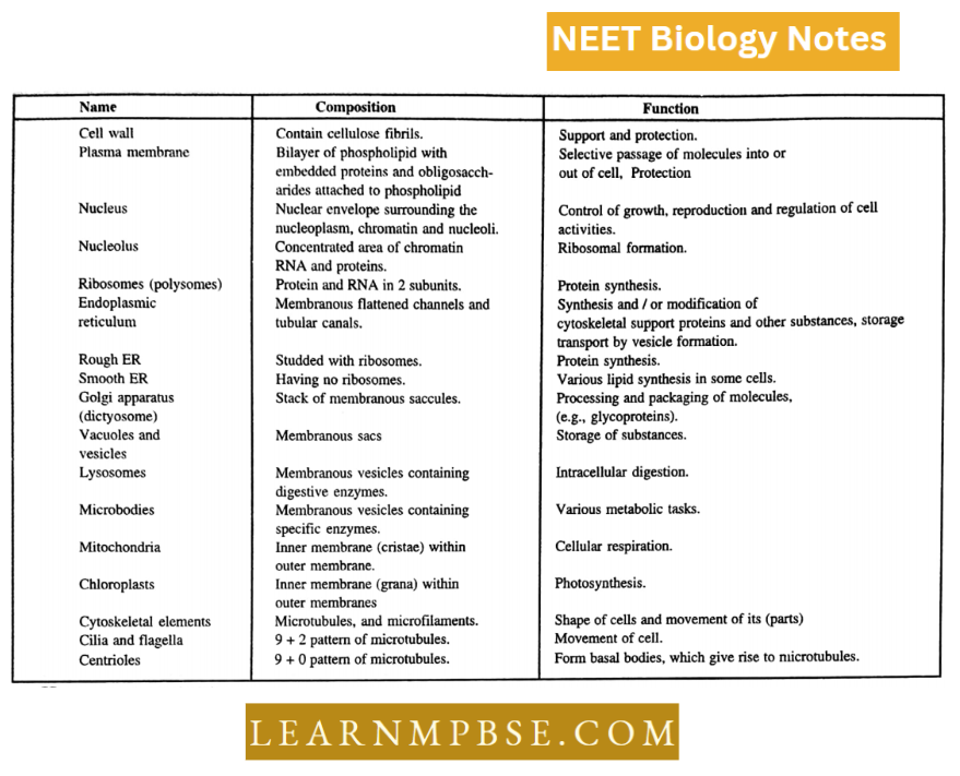
NEET Biology Cell Unit Of Life Cell Organelles
Cell organelles are of the following types depending upon several membranes.
- Single unit membrane-bounded organelles. Microbodies (Peroxisomes, Glyoxysomes, Sphaerosomes, Lysosomes, Golgi bodies, ER, vacuoles.)
- Double unit membrane-bounded organelles. Plastids, Mitochondria and Nucleus.
- Amembranous organelles: Ribosomes, centrioles, spindle, microfilaments and microtubules.
Endoplasmic Reticulum
The endoplasmic reticulum [ER] consists of an interconnected system of membrane-bound channels in the cytoplasm. It forms 30 to 60% of the total membranes of eukaryotic cells.
Electron microscopic observations by Porter, Claude and Fullam (1945) revealed a network of delicate strands and vesicles in the cytoplasm. The term endoplasmic reticulum was first used by Porter and Kallman (1952).
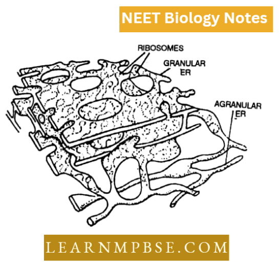
The endoplasmic reticulum consists of cisternae, tubules and vesicles. It occupies 10% of the total cell volume. The cisternae are broad, flat, membrane-bound spaces arranged parallel to each other to form lamellae.
They are interconnected with each other. The tubules are 50 -100 A in diameter and appear as circles in sections. The vesicles or sacs appear as membrane-bound isolated globular cavities.
There are two morphological types of the ER. When some elements of ER are studded with small granules called ribosomes, it is called rough or granular ER. The other type is smooth or agranular, marked by the absence of these granules.
The strands of HR consist of biomembranes formed of lipoprotein and the space between them varies. SER in muscles is called sarcoplasmic reticulum and myeloid bodies in pigmented epithelial cells and retina of the eye. Ergnstoplusm is the basophilic region of cytoplasm containing RER. Desmotubiiles arc extensions of ER through plasmodesmata.
Cell The Unit Of Life NEET Notes
Functions. The ER and nuclear membrane arc are directly connected. The ER is connected with intercellular transport and cellular metabolism. ER also plays an important role in the synthesis of nuclear membranes during cell division. It also helps in protein synthesis, glycogen synthesis and storage, lipid synthesis and storage and formation of microbodies.
The ER gives additional mechanical support to the colloidal structure of cytoplasm and increases surface area for absorption.
Synthesis of steroids and hormones and the formation of visual pigments from vitamin A are also associated with ER. It synthesizes ascorbic acid and also takes part in detoxification of toxic chemicals.
NEET Biology Cell Unit Of Life Ribosomes
Ribosomes. Ribosomes are nucleo-protein protoplasmic subspherical structures without a covering membrane, negatively charged with a diameter of 150 to 250 A meant for a polypeptide or protein synthesis.
They were discovered by Palade and thus called “Palade particles”. These arc smallest ameinbranous cell organelles. Later these were named ribosomes. These are present in cytoplasm in the form of groups, called polysomes. Ribosomes are made up of ribonucleoprotein.
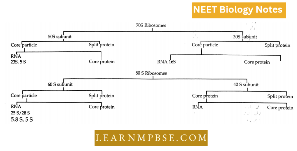
Ribosomes are of two basic types: 70S ribosomes in Bacteria and Blue-green algae, mitochondria and chloroplast, and 80S ribosomes in Eukaryotes. A 70 S ribosome has three molecules of r RNA 16 S, 5 S, 23 S, and 50 proteins mols.
80 S ribosome has four mols of rRNA (5 S, 5.8 S, 18 S and 28 S) and 80 mols of proteins. The 70S ribosomes are inactivated by the antibiotic Chloromycetin. The 80 S ribosomes are inactivated by cyclohexamide.
Ribosomes 80 S contain 40% and 60% RNA and protein respectively. Ribosomes are the seats of protein synthesis.
In mammalian mitochondria, ribosomes are of 55 S type. Proteins of all ribosomes are similar but it is the RNA which differs from ribosome to ribosome.
Prokaryotic And Eukaryotic Cells Differences NEET
Ergosomes or Polyribosomes (Rich 1963) are formed by protein synthesis.
- A tenfold increase in Mg2+ cone unites ribosomes to form Diamers. 70 S ribo some form 100 S while 80 S forms 120 S. If cone, is decreased by 10 folds, the ribosomes disassociate. Only unemployed ribosomes disassociate while active or those involved in protein synthesis do not disassociate.
- Lnke in 1976 proposed an asymmetrical model whereas Stefflar and Whittman, 1977, proposed a quasisymmetrical model for ribosomes.
- About 75 % of the ribosomes of cells remain bound to the C-face of the E.R. membrane through a glycoprotein ribophorin. In the case of He La cells the number of ribosomes is 15%.
- Proteins of all ribosomes are similar, however, it is r RNA that differs from one ribosome to another ribosome.
- Proteins synthesized on free ribosomes are used within the cell and those synthesized on bound ribosomes are used outside the cell.
Golgi Complex
Golgi Complex. These structures are also known as dictyosomes, gold some, idiosomes, Golgi apparatus or hypochondria, Dalton’s complex or Baker’s body.
In 1898, an Italian neurologist C. Golgi described certain previously unknown bodies in the cytoplasm of nerve cells of bam owls and cats using a special stain, Earlier the Golgi complex was thought to be present only in animal cells but recent research has shown that is present in plant cells.
Dalton and Felix observed them under TEM
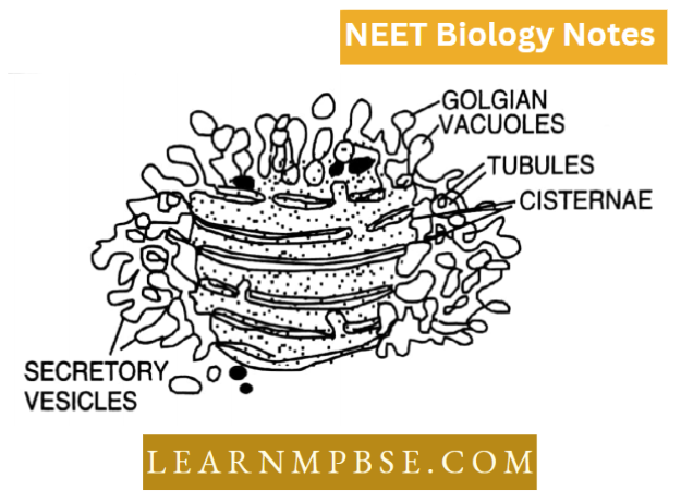
Three distinct components are visible in the Golgi complex, they are
Tubularorflattened fluid-filled sacs or cisternae, each is membranous and parallel or concentrically with a convex (forming) surface towards the plasma membrane and the opposite concave surface is termed maturing surface,
Transition vesicles are small drop-like structures Secretory vesicles are present on the sides and the maturing face of Golgi.
NEET Biology Cell Structure Notes
The elements of the Golgi complex may arise from ER, nuclear membrane or the pre-existing dictyosomes. It contains lipids, proteins and glycoproteins. Phosphatases, RUA, Phospholipids, ATP ases and S-nucleotidases are also present.
The metallic impregnation technique (osmium chloride silver salts)
NEET Biology Cell Unit Of Life Mitochondria
Mitochondria (Mito = thread, chondrion = granule) were discovered by Kolliker (1850) and the name mitochondria was given by Benda. Altman (1886) called them as ‘bioplasts’. He considered them as symbionts comparable to bacteria. In plants, mitochondria were discovered by Meaves (1904) in Nymphaea.
Mitochondria are also called Chondriosomes or Chondriomites plastochondria or filavermicules or bioplasts.
Mitochondria occur in the cytoplasm of plants and animal cells, the average length of the mitochondrion is 3 to 5 microns and the average diameter is 0.5 to 1.3 microns. They can be stained with Janus green B.
Using ultra vibrations and detergent actions, two membranes of mitochondria can be separated to study their structure. The mitochondrion without an outer membrane is called a mitoplast.
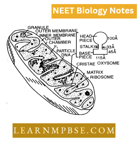
The mitochondrion is bounded by two membranes, the outer membrane and the inner membrane.
There are two chambers. The space between the two membranes is called the outer chamber and the space bounded by the inner membrane is called the inner chamber. The inner membrane forms mitochondrial cristae.
The enzymes of the Krebs cycle, flavo-proteins and cytochromes are present in mitochondria which are the centre of respiration. Mitochondria are also involved in oxidative phosphorylation and ATP synthesis. These are also called as ‘Power house’ of the cell.
Mitochondria arise from pre-existing mitochondria or ER. Mitochondria are semi-autonomous bodies. Nass, Nass and Afzeluis (1965) have shown the presence of DNA in mitochondria (MDNA). It constitutes 17c of the total DNA of the cell.
It is a double double-stranded, naked, granular, slim, long molecule with a higher G-C ratio. It is a circular molecule in most higher animal cells but is linear in several eukaryotic plant cells. Ribosomes arc of 70S type (55S ribosome in mitochondria of mammals) RNA and 70 types of enzymes are present.
Cell Organelles Structure And Functions NEET Biology
The mitochondria of whole cells are collectively called chondriosome or chondriomes. On cristae are present oxysomes or F0—Fj particles which are tennis racket-shaped bodies.
These particles were discovered by H.F. Moran and Chanre. These are also called as ETS particles as electron transport systems and ATP synthesis occurs on them. Besides this, mitochondria are concerned with lipid synthesis and elongation of fatty acids.
In prokaryotes where mitochondria are absent, the site for oxidative phosphorylation and electron transport, including dehydrogenases is the plasma membrane.
Single mitochondrion occurs in the case of:
- Microsterias Trypanosoma Chlorella.
- The maximum number is 5,00,000/cell in flight muscles.
- Meaves first to observe mitochondria in plants (Nymphaea).
- They can be stained by Gentian Violet and Janus green B.
- Cytochrome oxidase is present in the inner mitochondrial membrane.
- Mitochondria are rich in Mn. They are yellow due to riboflavin. They live for 5×10 days membrane only
- In all probability, the mitochondria arise from the nuclear envelope by evagination or by division of pre-existing mitochondria.
- Pyruvic acid enters mitochondria and is converted to Co. A in per mitochondrial space.
- ATP molecules are synthesised in the head.
- There are 5 chemical complexes. The first four constitute the electron transport system and the fifth transfers and conserves energy and (helps in) ATP synthesis.
- Mobile carriers = CoQ and Cyt C.
- Flavoproteins are located in Complex 1
NEET Biology Cell Unit Of Life Lysosomes
Lysosomes are diminutive vesicles encased by a singular membrane that contain hydrolytic enzymes in crystalline or semicrystalline forms.
- Christian de Duve discovered them. Lysosomes are present in nearly all animal cells, with the exception of mammalian red blood cells.
- Their diameters are 0.4 and 0.8 ™. These are lacking in plant cells; nevertheless, P. Matile (1964) has identified them in the fungus Neurospora.
- They are also referred to as ‘Suicidal bags’ or ‘Atom bombs’ due to their containment of hydrolytic enzymes.
- Lysosomes are most abundant in white blood cells, macrophages, osteoclasts, and similar cell types.
- Plant cells are devoid of lysosomes, with the exceptions of Neurospora, maize root tips, yeast, and the seeds of peas and cotton. Lysosomes originate from the M face of the Golgi apparatus.
- Lysosomes execute various duties, including the digestion of food, the phagocytosis of foreign particles, involvement in metamorphosis, scavenging, supplying enzymes for the degradation of egg membranes, and facilitating the disintegration of ageing and dead cells.
- Lysosomes eliminate toxins through engulfment and appear to be crucial for cellular division.
- Additionally, they have an inherent mechanism that allows a cell to adapt.
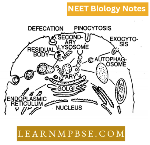
Lysosomes exist in more than one morphological form, hence show polymorphism and they are of the following four types:
- Primary lysosomes (also known as original lysosomes storage granules or inactive lysosomes).
- Secondary lysosomes (heterolysosome or phagolysosome heterophagic vacuoles or digestive vacuoles).
- Autophagic vacuoles (autolysosomes or autolysosomes).
- Residual bodies (telo lysosomes dense bodies or tertiary lysosomes).
NEET Biology Cell Unit Of Life Plastids
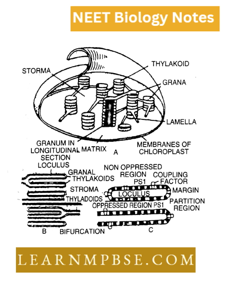
Plastids. Plastids (Haeckel, 1866) are semi-autonomous organelles having DNA and a double membrane; envelope which synthesizes or stores different types of organic compounds.
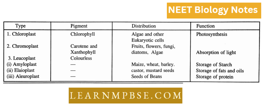
The chloroplasts are green plastids (Schimper 1833) which take part in the synthesis of organic food from inorganic raw materials in the presence of radiant energy or light and the process is termed photosynthesis.
Eukaryotic plants contain a heterogeneous group of cell organelles called plastids which vary in morphology and function, some plastids are colourless, called leucoplasts. They store reserve materials, such as starch, proteins and lipids.
Green plastids are called chloroplasts which contain chlorophyll. Still others the chromoplasts. contain carotenoids which are brilliant red or yellow.
All three plastids differentiate from small spherical amoeboid structures 0.4 to 0.9 um in diameter called proplastids. Plastids are the largest cell organelles.
The increasing order of plastid size is as follows:
Chloroplast → chromoplast → elaioplast → aleuroplast → amyloplasts.
The distribution of pigment systems are coupling factor in thylakoid membranes. The chloroplasts are usually discoidal or lens-shaped double membrane organelles.
They are made up of chlorophyll a and b carotenoids, lipids, proteins and nucleic acid. DNA in chloroplast was reported by Ris and Plaut.
The chloroplasts are ‘autonomous bodies and. thus, are also called “cells within cell organelle.” The membrane-bound matrix of chloroplast is called stroma where dark reaction or Calvin cycle of photosynthesis takes place. In the stroma are present thylakoids which form grana acting as site of light reaction where assimilatory power (ATP 4- NADPH,) are synthesized.
Fluid Mosaic Model Of Cell Membrane NEET Study Material
The smallest and most functional unit of chloroplast is called the photosynthetic unit or Quantosome. The term Quantosome was given by Park and Biggins (1964).
Each Quantosome consists of 250 molecules ofchlorophyll. The molecular weight quantosome is 2 x 10⁶.
The size ofquantosome is length = 1 80 A width = 155 A and thickness = 100 A. It is considered that chloroplasts might have originated from a symbiotic relationship between an autotrophic microorganism capable of transforming energy from light and a heterotrophic cell.
- Thylakoid membrane consists of 2 photosystem complexes namely photosystem 1 and photosystem 2 their reaction centres being P-700 and P-680.
- LHCP = Light-harvesting complex protein. It is present in the thylakoid membrane and is photochemically inactive.
- Plastids are called intracellular parasites and a symbiont hypothesis is used to explain them.
- The plastidial system carrying genetic information is called the plastidome.
- Chloroplast of higher plants chemically is composed of
- Protein: 33-35%
- Chlorophyll: 9%
- Lipids: 20-30%
- Carotenoids: 4-5%
- Nucleic acids : 3-4%, traces of Vit K and A, Ribosomes 70
- Protein lipid ratio in the chloroplast is 40: 30
- No. of grana in chloroplast ranges from 10-100
- C4 plants have dimorphic chloroplasts.
- Chloroplasts move in the cell due to cyclosis.
- Chloroplast is more important than chlorophyll as carotenoids in chloroplast protect chlorophyll from photo-oxidation.
- Chloroplasts with nitrogen-fixing genes are called nitroplasts.
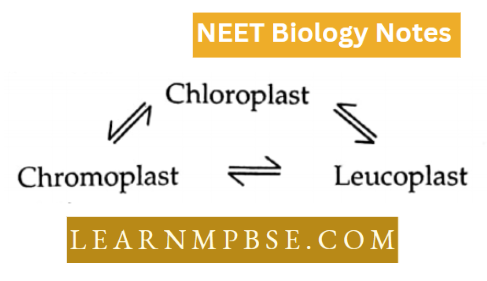
- The three kinds of plastids are interconvertible
Classification based on pigmentation.
- Chromoplast: coloured plastids
- Chloroplast: ‘Chlorophyll a & b c.g. green algae
- Phaeoplast: Fucoxanthin Example brown algae, diatoms.
- Rhodoplast: phycoerythrin Example red algae
- Blue-green chromoplast, ‘phycocyanin Example Blue-green. Algae
- Leucoplast colourless plastids
- Amyloplast: Starch storing
- Elaioplast (Oleasome): Fats storing.
- Aleuroplast (Proteinoplasts) : protein storing.
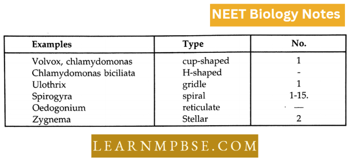
Shapes
- The chlorophyll in grana is surrounded by 2 protein layers (Y-shaped) in between the protein and chlorophyll there occurs lipid chlorophyll.
- Leaf palisade mesophyll cells of higher plants contain 20-40 chloroplasts.
- Single thylakoid occurs in the granum of Rhodophyceae.
- Quantosome is believed to have (160 a + 70 b) chlorophyll and 50 carotenoid molecules.
- Chloroplasts which lack granum are often described as agranal or Kranz type.
NEET Biology Cell Unit Of Life Microbodies
Peroxisomes (Uricosomes)
These are single membrane-bound vesicles with a size ranging from 0.5 to lim. They were discovered by Tolbert and the term peroxisome was coined by de Duve (1969) for those microbodies which are rich in enzymes peroxidase classes and catalases and produce hydrogen peroxide during their degrading activity.
These vesicles contain fine, granular substances that may condense to form a central core or nucleoid.
The peroxisomes without nucleoids are called micro-peroxisomes. The enzymes present are:
- Uric acid oxidase
- α – amino acid oxidase.
- α – Hydro xylic acid oxidase.
- β Hydroxylic acid oxidase.
- NADH-glyoxalate reductase.
- NADH-isocitrate dehydrogenase.
- Catalase.
- Peroxidase.
- Peroxisomes generally arise from ER. These bodies take part in H202 metabolism and in green plants, they carry out photorespiration.
Sphaerosomes
Hanstein (1880) observed small highly refractive bodies of a denser substance in the cytoplasm of plant cells and later Danglard termed them sphaerosomes. They are microbodies which take part in the storage and synthesis of fats.
They were first discovered by Perner in 1953. Sphaerosomes are small spherical and refractile vesicles which are 0.5— 1.0 mm in diameter.
They arise from the ER and are surrounded by a single membrane. 98% of a phagosome is fat. Proteins constitute the remaining 2%. Some proteins are probably enzymatic and take part in the synthesis of fats.
Fluid Mosaic Model Of Cell Membrane NEET Study Material
Because of the presence of fat, phagosomes can be seen under light microscope after staining the cells with Sudan dyes and osmium tetraoxide. Sphaerosomes occur abundantly in the endospermic cells of oil seeds.
Sphaerosomes of some tissues Example tobacco endosperm, and maize root tips contain hydrolytic enzymes. Therefore, they are considered to have lysosomic activity.
Glyoxysomes
- They are microbodies which contain enzymes [5-oxidation of fatty acids and glyoxalate pathway. These microbodies were discovered by Breidenback and Beevers (1967) from the extracts of the endosperm of germinating castor beans.
- They are also present in the cells of some fungi. Like other microbodies, glyoxysomes have a single covering membrane and a matrix with a crystalloid core.
- The matrix contains enzymes for (3-oxidation of fatty acids to produce Acetyl CoA. The latter is metabolised in the glyoxylate cycle to produce carbohydrates.
Lomasomes
They were discovered by Moore and McAear (1961) and are often referred to as border bodies. These bodies are present between the cell wall and cell membrane of both lower and higher plants. They play a role in secretion, increasing the surface area for the diffusion of substances involved in cell wall formation or breakdown in membrane proliferation and endocytosis.
Transosoines
- These bodies were discovered by Press (1964) and are so far known to occur only in the ovarian follicles of birds.
- These are circular and consist of triple-bonded envelopes with several ribosome-like granules attached by short stalks to the innermost band. It is believed that they have a role with the developing yolk granules but their exact function is not known.
NEET Biology Cell Unit Of Life Centrioles, Cilia And Flagella
Centrioles. They were first observed by Van Benden in animal cells. They are minute microtubular rods which can help to form their duplicates without having DNA and membranous covering.
Usually, two centrioles called diplosomes are present near the nucleus of all eukaryotic animal cells, primitive plants and all eukaryotic where flagellate structures and all eukaryotic plants where flagellate structures are present.
Mature centrioles are 0.2 lim in diameter and vary in length from 0.2 to 0.5 pm. A pair of centrioles surrounded by a clear area of cytoplasm, the centrosphere, is called a centrosome. Each centriole consists of nine sets of microtubules arranged circularly.
These tubules are equally spaced. Each microtubule is a triplet (diameter 250 A) of microtubules or subfibres embedded in a matrix designated as A, B, C and from inner to outer subfibre. Each subfibre is made up of 11—13 protofilaments of tubulin. The A-subfibre of a triplet is attached to the C-subfibre of the adjacent triplet by strands called linkers.
The central hub is (20 A in diameter), and from it arises 9 proteinaceous connectives called spokes, one towards subfibre.
NEET Biology Cell Structure Notes
A thickening ‘X’ is present at the outer end of each spoke and another Y is present between the two X thickening and are connected.
There are dense, amorphous, protoplasmic structures called macule satellites or pericentriolar structures on the surface of centrioles. Centrioles are locomotory structures and their role in cell division is acquired secondarily.
Cilia and flagella
- (Studied by Englemann and confirmed by Jensen 1881) are hair-like protoplasmic processes of cells capable of undergoing movements and thus create a current jn -any fluid for locomotion and passage substances.
- Both cilia and flagella have the same basic structure and differ in number, size and type of movement.
- Chemically, they are mainly formed of protein and lipids, traces of carbohydrates and nucleotides are also present. Tubulin protein occurs in peripheral fibrils while dynein is found in the side arms of A-subfibrils spokes and central fibrils.
Structure
- The cilia and flagella are essentially of the same structure and both arise from basal bodies. Cilium is surrounded by a membranous covering which is an extension of the plasma membrane. Both the membranes are separated by a space of 90 A.
- The space between the limiting membranes of the cilium is filled with a matrix. In the matrix is embedded axial fibril complex.
- The central axial filament complex consists of eleven microtubules arranged in two radii; out ofthese nine are double and are situated at the periphery and two single microtubules are placed in the centre.
- Each of the nine outer tubules is 360 A in diameter and composed of two subunits. A sheath surrounds the central fibrils. The outer fibrils bear a pair of arms Radial lamellae, each with a thin secondary fibril, occur between central and outer fibrils.
- Central singlet fibres are made up of a protein dynein with ATP-ase activity. Outer double fibres are made up of tubulin protein while A-B linkers are composed of protein nexin.
NEET Biology Cell Unit Of Life Cytoskelclal Supporting Structures
- Intermediate filaments are long, unbranched having structure intermediate between microtubules and microfilaments and consist of a head, central rod and tail. First discovered in muscle cells (1968).
- Microtubules earlier called neurotubules were first discovered by Sabatini, Bensch and Barnette (1963).
- Microtubules and microfilaments form a complex structural framework of the cell. They are concerned with movement by or within the cell. They also can maintain the shape of the cell.
- Microfilaments are long, thin, cylindrical rods composed of actin (protein).
- Microfilaments are 6-10 nm in diameter and contract with the help of myosin.
- Microfilaments may extend into the cytoplasmic core of microvilli.
- Microfilaments are responsible for movement of the plasma membrane during cell mobility and endocytosis; contraction of muscle fibres and movement of microvilli.
- Microtubules are elongated unbranched, cylindrical tubes of about 25 nm in diameter.
- Each tubule encloses a light central core of 15 nm diameter. The microtubule’s wall is made up of 13 longitudinal filaments, each is a polymer chain of tubulin protein.
- Microtubules occur singly or in bundles in cytoplasm radiating from the controls to the peripheral parts.
- These form the skeleton of cilia and flagella and spindle during cell division helping in the movement of chromosomes. They can be broken down and reassembled in another part of the cell.
NEET Biology Cell Unit Of Life Interphase Nucleus
The nucleus of the cell which is not in the process of division is called interphase nucleus.
Robert Brown first noticed the largest organelle, the nucleus. It is present in all eukaryotic cells except RBC. Cells usually have one nucleus (mononucleate), but some cells may be binucleate or polynucleate.
It may be placed in the centre or one side of the cell. It is mostly spherical or oval in shape or it may be elongated or even lobulated. The size of the nucleus depends upon its proteins and DNA contents.
The cytoplasmic and nuclear masses remain in the state of equilibrium known as endoplasmic or kanophismic index (NP or KPL)
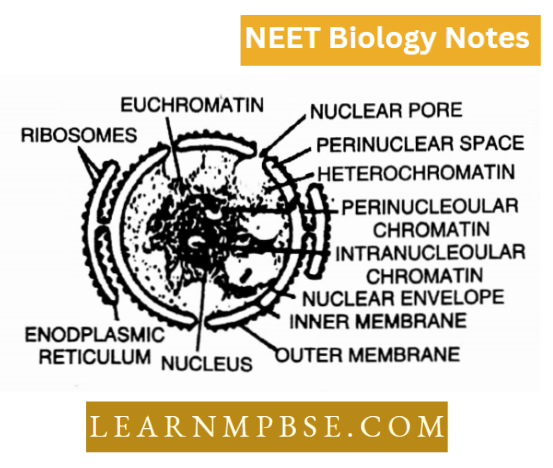
\(\mathrm{NP}=\frac{\text { Volume of nucleus }}{\text { Volume of cell }- \text { Volume of nucleus }}\) \(=\frac{\text { Volume of Nucleus }}{\text { Volume of eytoplasm }}\)Nuclear envelope. The nucleus is bounded externally by a double membrane, the nuclear membrane, which beats sub-microscopic pores and through these pores it is in continuation with the membranes of the endoplasmic reticulum.
The space between the two nuclear membranes is called the perinuclear space. Each membrane is 75 A in diameter and perinuclear space is 150-350 A wide.
In structure and function, the nuclear membrane resembles the cell membrane. It disappears during cell division and is redeveloped tram the membrane of the endoplasmic reticulum. Internally, the nucleus is filled with a viscous fluid, the nuclear sap. nucleoplasm or karyolymph. It is rich in protein and contains nucleic acid.
Nucleolus. (PI. Nucleoli Fontana 1784, discovered which was described by Wagner (1840) and Bowman (1S40) provided the present name. Nucleolus is an irregularly shaped often dense and compact body. It consists of protein and RNA. The patient forms SOT of the dry weight of the nucleolus. The nucleolus performs three functions, synthesis of proteins.
Mitochondria And Chloroplasts Roles NEET Exam Preparation
synthesis of ribosomal RNA and transfer of genetic information from the nucleus to the cytoplasm. It is interesting to note that the nucleolus disappears during cell division. Chromatin material or network is a granular mass which lies in the nucleoplasm.
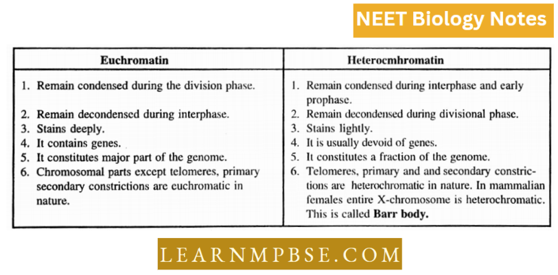
The chromatin with fine threads is called euchromatin. The chromatin material consists of DNA. RNA. histone and acidic protein. During cell division, the chromatin material organizes itself and becomes distinct as chromosomes.
Differences Between Euchromatin And Heterochromatin
Nuclear functions. Robert Brown (1831) discovered the nucleus which was later mentioned as store storehouse of heredity information. Van Benedan and Hertwig demonstrated the role of the nucleus during the fusion of male and female gametes.
Boveri (1889) experimented with sea urchin eggs and established that survival, growth and reproduction are controlled by the nucleus.
Hammerling’s (1913) grafting experiment with Acetabularia Mediterranean proved that morphology is governed by the nucleus.
Hofmeister observed chromatin threads during cell division. Sutton and Boveri observed parallel behaviour between chromosomes and genetic characters and proposed the chromosome theory of heredity.
The DNA constituents of the nucleus act as the chemical basis of genetics. Structure of chromosome.
The chromatin network undergoes condensation, and coiling and is transformed into a definite rod-shaped structure of the chromosome. The chromosomes were discovered first by Hofmeister and the word chromosome was given by Waldeyer (1888).
A typical chromosome has the following parts:
Pellicle. It is the outermost covering membrane chromosome. However, Ris (1945) denies the presence of pellicles.
Matrix. It is the ground substance of chromosomes and contains a chromatin network.
Chromonema. There are two spirally coiled threads in the matrix, known as chromonemata. They are seen throughout the length of the chromosome. Each chromonema is 800 A in thickness and consists of one molecule.
Chromoniercs. These are dot-like or hcad-like structures of chromonema which have coiled up in specific ways to form complex and dense chromatin, the chroinomcres.
Prokaryotic And Eukaryotic Cells NEET
Centromere. It is situated in the region of primary constriction. In this region, the two chromonema join to form one.
The position of the centromere helps in the identification of chromosomes. According to the position of the centromere the chromosomes are of four types: metacentric, submetacentric, acrocentric and telocentric.
According to the number and presence or absence of centromeres, chromosomes arc of the following types:
Acentric (without centromere), monocentric, dicentric and polycentric with one, two or many centromeres.
Secondary Constriction. This constriction may be present in one or both arms of the chromosomes. This is the place where a nucleolus appears and disappears during the cell cycle. Thus it is also known as the nucleolar organizing region.
Satellite. The distal spherical part of chromosome above sec. constriction is known as a satellite. The chromosomes having satellite arc are known as SAT-chromosomes where SAT stands for Sine Acid Thymunucleinico.
Telomere. Each chromosome has polarity. Its terminal ends are physiologically different from the rest of the chromosomes and are known as telomeres. They avoid sticking of chromosomes in the nucleus. If by X-rays these ends arc broken, then one chromosome will stick to another chromosome.
In chromosomes, DNA and histones arc bonded to form dcoxyri bonuclcoprotcin. The most accepted model for the arrangement of DNA and histone protein is the nucleosome or sole noid model (Woodcock).
The term nucleosome was given by. Outlet ( 1 975). To accommodate the nucleosome model for DNA, Crick and Watson proposed the Kinky helix model for DNA. A nucleosome is a repeating unit of chromatin (125 nm is diameter = 200 base pairs + 2 molecules each of H2A, H2B, H3 and H4)
Lampbrusli Chromosomes. Such chromosomes are found in the oocytes of amphibians and appear like a brush which was used in the olden times to clean street lamps. That is why, they are known as lampbrush chromosomes.
A lampbrush chromosome consists of a central main axis made up of DNA whereas the matrix which consists of RNA and protein is present in the form of a loop of the main axis. The loops lie opposite to each other.
Prokaryotic And Eukaryotic Cells NEET
Each loop consists of an axial fibre which is covered with a matrix. When it is treated with ribonucleases (an enzyme which breaks down DNA) it is broken down indicating that it consists of DNA.
When it is treated with ribonucleic (an enzyme which breaks down RNA), trypsin and pepsin the matrix of the loop is removed. From that, it can be concluded that the matrix is composed of RNA and protein whereas the axial fibre of DNA.
Polytene Chromosomes. They were first observed by Italbiani (1881) in the salivary gland of Chirommous and hence sometimes are also known as salivary gland chromosomes.
They are also found in Drosophila. In Chironomus, usually, one somatic cell consists of 8 polytene chromosomes. They measure 1000 times larger than normal chromosomes. They are 2,000 Im in length.
A polytene chromosome consists of several parallel strands, chromonema. The coils containing polytene chromosomes continue to grow without undergoing mitosis. One chromosome may have as many as 1024 chromonemata. All strands are genetically identical.
- m-chromosomes. Minute-sized (less than 0.5 mm) chromosomes are found in many bryophytes. and insects.
- Chemically, Chromosomc is formed of 40% DNA, 50% histones, 1.5% RNA, etc. H, H2A and Hyn proteins are lysine-rich (2 is very lysine-rich) while H3 and H4 are arginine >ric)i polypeptide chains.
- Satellite is also called Trabant.
- NOR constitutes 03% of total nuclear DNA. NOR is also called secondary constriction I.
- Muller (1938). Coined the term telomeres.
- Vejdovsky (1912). Coined the term chromonema.
- Du Praw : (1966). Proposed uncinematic folded fibre model of the chromosome.
- Finch and Klug (1976). Proposed solenoid model. It states that DNA is associated with histone proteins to form a series of beads called nucleosomes.
- Nucleosomes: Fundamental packing unit of chromonema.
NEET Biology Cell Unit Of Life Cell Inclusions
Reserve food. It may be in the form of starch grains (Example in potato-tuber cells) or glycogen granules (Example in liver and muscle cells of animals), protein granules (Example in aleuroplasts of the endosperm of maize seed), or oil droplets (Example in adipocytes of fat bodies; endospermic cells of castor and coconut and in cotyledon cells of groundnut and mustard seeds).
Gases. These include O2, CO2, N2 etc.
Inorganic crystals. These include crystals of calcium salts and silica. Silica is generally found in the epidermal cells of grasses. Crystals of calcium carbonate, called cystoliths, are found in the epidermal cells of Mimordica (Kerala), while crystals of calcium oxalate are called raphides and are found in the epidermal cells of Eichhomia (water hyacinth). Hyaloplasm also contains organic acids like tartaric acid (In tamarind), citric acid (citrus fruits) and malic acid (as in apples).
Excretory/Secretory Products. They include mucus, tannins, resins, gums, alkaloids, latex, etc.
Latex. It is a crystallo-colloid fluid secreted by latex tubes or Iaticifers of two types, latex cells (non-articulated Iaticifers, Example Banyan, Calotropis, Oleander) and latex vessels (articulated Iaticifers, Example Poppy, Rubber plant, Sonchus). Latex can be watery (For example Banana), milky (For example Banyan) or coloured (For example Poppy).
The latex of Hevea brasiliensis yields rubber, that of Poppy forms opium while the latex of Papaya contains the protein-digesting enzyme papain,
Gums. They are degradation products of cell walls, for example, gum Arabic (Acacia Senegal).
Gum-Resin. It is mixture of gum and resin, Example root of Ferula asafoetida (asafoetida).
Resins. They are acidic oxidation products of essential oils which are insoluble in water but soluble in alcohol/ turpentine. An example of hard resin is shellac. Pine resin and Canada Balsam are oleo-resins (resins with associated essential oils).
NEET Biology Important Notes PDF
Tannins. They are astringent, acidic, phenolic compounds, related to glucosides, found in leaves (For example Tea), bark (For example Acacia nilotica, Walnut or Juglans regia) fruit (For example Caesalpinia, Betel Nut), Dyes related to tannins are cutch (heartwood of Acacia catechu), and haematoxylin (heartwood of Haematoxylon).
Alkaloids. They are bitter nitrogenous by-products, often poisonous and with medicinal properties, Example quinine (bark of Cinchona officinalis), atropine (leaves and tops of Atropa belladonna), nicotine (leaves of Nicotiana tabacum (tobacco), morphine (latex of Papaver somniferum (poppy), reserpine (roots of Rauwolfia serpentina), colchicine (corms of Colchicum autumnale), thein (Tea leaves),
Glucosides present is crucifers. They are aromatic compounds having glucose/carbohydrates, for example, saponin, digitoxin, digitalin, and amygdalin. Many of them are medicinal, (viii) Essential Oils. They are volatile aromatic oils secreted by special glands, Example Lavender, Rosemary oil, Menthol, Eucalyptus oil,
Nectar. It is the sugary secretion of parts of flowers to attract insects and other animals for pollination. Nectar contains glucose,’ fructose and sucrose.
Pigments. These are found in special cells, called chromatophores, present in the skin (dermis) of vertebrates (fishes, amphibians and reptiles).
The chromatophores are of two types: melanophores have brown or black pigment, and lipophores contain red, yellow and orange pigments. Birds and mammals have a pigment in their feathers and hair. In the human skin, the pigment-containing cells occur in the deeper epidermal cells
- Prokaryotic cells are represented by bacteria, blue-green, algae, mycoplasma or PPLO, spirochaete and rickettsiae generally smaller than eukaryotic cells but multiply more rapidly.
- The bacterial flagellum is formed of filament, hook and basal body which is a hollow rigid, cylindrical filament longest (1-70 nm), formed of flagellin protein.
- The basal body of the bacterial flagellum is a complex part of the flagellum, consisting of four and two rings connected to the central rod in Gram-ve and Gram+ve bacteria respectively.
- The filament, hook and basal body are arranged in such a manner to permit the filament to rotate by 360° rather than undulating back and forth like a whip. In the plasma membrane of eukaryotic cells sterol molecules are more rigid than phospholipids and therefore the presence of sterol confers stability on each eukaryotic membrane.
- Three principal types of protein filament-microfilaments (7nm), microtubules (25nm) and intermediate filaments (8-10nm) are cytoskeletal elements which allow the eu¬ karyotic cell to adopt a variety of shapes and carry out directed movements.
- In Golgi bodies, materials are transported from the cis to the trans face by vesicles that keep budding off from the cisternal edge to the next cell and so on.
- The sequence of the size of the organelles is as follows:
- Lysosome(0-2-0-8pm) <Sphaerosome (0-5-1 -0pm) < Per peroxisome (0-5- M pm)< Ribosome (l-5-2-0pm)
- Pinosomes and phagosomes are collectively known as endosomes.
- The vacuoles of plant cells are bounded by a single semipermeable membrane called tonoplast, whereas the vacuoles, of animal cells are by, a lipoprotein membrane.
- Vacuoles are of four types depending upon function t. sap vacuoles, contractile vacuoles, food vacuoles and air, rapidly.
- vacuoles/ The proper folding of proteins following synthesis is assisted by a special protein called chaperones.
- Mitochondria can be seen in inactive or orthodox state and active or condensed state.
- Both mitochondrial (mt DNA) and chloroplast DNA (cp DNA) containing limited genetic information are circular although cp DNA is much bigger than mt DNA
- The amount of DNA is low in a chloroplast i.e. 10-15 to 10y grams per chloroplast or 0-03% of its dry weight.
- Cilia and flagella having a 9+2 arrangement are associated with the motility of cells.
- Karyotype. The arrangement of chromosomes in terms of their relative size, form and number is called karyotype. It consists of the arm ratio and position of the centromere also. It is characteristic of a species.
- Karyotype preparation can be done by either banding technique or fluorescence in situ hybridisation (FISH) and multicolour fluorescence in situ hybridisation (FISH) or Flow cytometry.
- Idiogram. A pencil sketch or photograph of the karyotype is called idiogram.
- Human Karyotype. Somatic cells contain a total of 46 chromosomes (Tijio and Levan 1956) or 23 pairs.
- Out of these 22 pairs are autosomes, one pair of sex chromosomes XX in females (homomorphic) and XY in males (heteromorphic).
Quanta to memory
- The term ‘cell’’ coined by Hooke (1665), is; a misnomer as the cell is not a hollow structure.
- Cytoplasm coagulates at temperatures above 60° C.
- Swammerdam was the first to describe the cell (RBC of frog).
- Largest cell—Egg of ostrich (6 inches in diameter with shell and 3″ without shell.)
- The nerve cell is the longest in the human body. The smallest cell (Mycoplasma gallisepticum—PPLO) measures 0.1 to 0.3 pm. Unicellular eukaryotes are 1 to 1000. pip in size. The size of human RBC is 7-8 pm.
- Cells of multicellular eukaryotes are 5 to 1000 pm in size.
- Fibre of Boehmeria (longest plant cell is 55 cm in size).
- PPLO (Mycoplasma Lauderhill) are also called “Jokers
of plant kingdom. - Transosome organelles are bounded by triple membranes.
- Organelles without membranes are ribosomes, centrioles,
microfilaments and microtubules - In human beings, cells of the kidney are the smallest and of nerve fibres largest.
- The size of human RBC is 7-8 pm.
- Cells of multicellular eukaryotes is 5 to 1000 pm in
size. - Fibre ofBoehmeria (longest plant cell is 55 cm in size). PPLO (Mycoplasma laiderwili) are also called “Jokers of the plant kingdom.”
- Transosome organelles are bounded by triple membranes. Organelles without membrane are ribosomes, centrioles, microfilaments and microtubules.
- In human beings, the cells of the kidney are the smallest and of nerve fibre largest.
- Intracellular compartments in cells help in the efficient functioning of cells.
- A pyrenoid is a proteinaceous body around which starch is stored in green algae.
- Protochlorophyll differs from chlorophyll in lacking 2 hydrogen atoms in two of its pyrrole units.
- Microtubules are involved in cell division.
- Two genome types are present in typical green plants.
- Basophilic ergastroplasm indicates the presence of ribosomes.
- Chromosomes are stained with acetocarmine processing or Acid Fuschia.
- Bioblast = Mitochondrion = Bioplast = unit of protoplasm capable of reproducing itself.
- The cell wall is capable of growth and is involved in many enzymatic activities, so is now considered as a living structure.
- A ripened fruit becomes soft because the pectates of the middle lamella solubilize on ripening.
- Myeloid bodies (granules at the base of retinal pigment cells) and Nissl granules (in the cyton of neurons) are RER (ER with ribosomes).
- Svedberg Unit (S). It is the rate of sedimentation or velocity of sedimenting the particles per unit of gravitational force, or lx 10-13 cm /sec/dyne/gm.
- The largest component of the cell is the nucleus while the smallest component is the microfilament.
- The largest cell organelle is the mitochondrion in animal cells and plastid in plant cells while the smallest cell organelle is the ribosome.
- The darkly staining property of chromatin is called heteropykinosis.
- Mitoribosomes of mammals have a sedimentation coefficient of 55 S.
- Smallest Human Cell. Erythrocytes with a diameter of 6-8 pm. Blood platelets are still smaller (2-3 pm in diameter) but they are considered to be cell fragments instead of cells themselves. (Malarial sporozoite is 2 pm in length).
- Coconut milk is widely used in tissue culture as it is rich in a kind of growth hormone named cytokinin.
- Callus and Cancer both are undifferentiated masses of cells.
- But calluses get differentiated into root and shoot by auxins and cytokinins in tissue culture and cancer cells fail to differentiate in tissue culture.
- The explant is an excised plant part used in tissue culture to
raise Callus. - Animal Cloning. Animal cells are totipotent like plant cells but there is difficulty in their dedifferentiation and later differentiation.
- Animal cloning has become possible by inserting the nucleus of a somatic cell into an enucleated egg and allowing the latter to develop into the uterus of a female. The first successfully cloned animal is Dolly, a sheep (1996—1997)