
Structural Organisation In Animals For NEET Biology Morphology Of Animals Pheretima Posthuma (Earthworm)
Earthworm belongs to the phylum annelida class oligochaeta.
Habitat:
Pheretima posthuma, the prevalent Indian earthworm, is a significant representative of the phylum Annelida. It is a fossorial creature.
- It inhabits burrows constructed in soft, damp soil abundant in decomposed organic material or humus.
- It excavates its burrows mostly in the pliable soils of gardens, lawns, or pastures. The burrows can stretch vertically downward to a depth of 30-60 cm.
- During spring and summer, the burrows may extend to a depth of 180-240 cm when moisture is present.
In the rainy season, the burrows become inundated, prompting the worms to exit and traverse the damp soil surface.
Structural Organisation In Animals NEET Notes
Read and Learn More NEET Biology Notes
Habits.
- Earthworms are nocturnal. During the daytime, it remains passively in the burrow with its anterior end directed upwards and posterior end downwards. At night, it becomes active.
- It may come out of the burrow and roam about. Sometimes it keeps the posterior end fixed to the burrow and moves the anterior part of the body in a circle. It feeds on soil particles rich in decaying organic matter or humus.
- It can also ingest directly the bits of leaves and other organic matter. The undigested food is egested as worm casting which appears as heaps on the soil.
- Earthworm respires through the skin which is very thin and highly vascular. It is always kept moist. The exchange of gases takes place by diffusion through the skin.
- Locomotion is affected by the contraction and relaxation of muscles of the body wall aided by setae or chaetae. The setae are embedded in setal sacs. They can be protruded or retracted. During locomotion, the anterior part of the body is extended.
- It is fixed to the substratum by setae. The posterior part of the body is drawn forward towards the anterior part. It is then fixed to the substratum by the setae and again the anterior part is stretched.
- This process is repeated. Thus, earthworms creep slowly on the surface of the soil. The setae hold the substratum firmly when the worm is making burrows, the earthworm can move backwards by reversing the direction of the setae.
- The backward movements occur when it has to come out of the burrow. The earthworm moves at the rate of 25 centimetres per minute.
- There are no special sense organs like eyes, ears etc. However the skin of earthworms lodges receptor cells and is highly sensitive to light and soil vibrations. The worm has a sense of smell and can detect food immediately.
- The earthworm is hermaphrodite bisexual or monoecious. It is oviparous. There is no self-fertilisation because it is protandrous. Thus, there is cross-fertilisation.
- During copulation, two earthworms are closely attached by their ventral surfaces in such a way that the head region of one is opposite the tail region of the other.
- There is a mutual exchange of spermatic fluid. Then the two worms separate.
Structural Organisation In Animals For NEET Biology Features Of Pheretima Posthima (Earthworm)
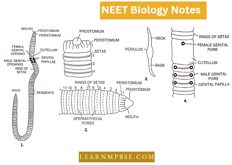
1. Earthworms are metamerically segmented nocturnal animals, that live in burrows made in the soft, moist soil of gardens, lawns, forests, pastures, flower beds and the soil rich in decaying organic matter or humus.
- They are rarely found in dry, sandy and clayey soils. They usually lie in the upper layers of soils.
2. The earthworm moves by muscular contraction and expansion of the body aided by chitinous setae present in the body wall.
3. Its body is elongated, 100-120 segmented, cylindrical and pointed at both ends. It measures about 150 mm in length and about 3 to 5 mm in diameter.
- The dorsal side of the earthworm is darker brown due to the deposition of a brown-coloured substance known as porphyrin which is derived from the decayed leaves, on which the earthworm feed.
- The first segment is called the peristome with an extension of the fleshy lobe, and proscenium, and segments 14-16 are covered by clitellum.
4. Apertures. Many apertures are found on the external surface which are openings of the coelom, alimentary canal, and the excretory and genital organs.
- Mouth. It is a crescentric anterior aperture present in the first segment called peristomium.
- Anus. It is a vertical slit-like aperture present at the posterior end of the last segment called the anal segment or pygidium.
- Female genital pore. It is a single median aperture of the oviducts present on the ventral side of the 14th segment. Eggs pass out of this aperture.
- Male genital pores. They are a pair of crescentric openings of the common prostate spermatic ducts present in the 18th segment on the ventral side.
- Genital papillae. There are two pairs of genital papillae present in the 17th and 19th segments on the ventral side. They bear the minute openings of accessory glands.
- Spermathecal pores. They are four pairs of apertures, each pair is present in the grooves of 5/6, 6/7, 7/8 and 8/9 segments. They are ventrolateral in position.
- Nephridiopores. These are minute openings of the integumentary nephridia present in the body walls. They are scattered in all the segments except the first two segments.
- Dorsal pores. These are minute pores of coelomic chambers located mid-dorsally, one in each inter-segmental groove, behind the 12th segment.
5. Body wall. It is covered with a very thick cuticle. The body wall consists of the following layers,
- Cuticle
- Epidermis
- Circular Muscles
- Longitudinal Muscles
- Coelomic Epithelium.
- The epidermis is one cell in thickness and consists of
- Mucus secreting cells.
- Albumen secreting cells.
- Sensory cells,
- Supporting cells.
- Basal Cells.
6. The Coelom. There is a true coelom present in earthworms. It is partitioned into so many coelomic compartments with the help of septa. The first septum lies between 4/5 segments and is thin and membranous.
Structural And Functional Organisation In Animals
- In the coelom is present milky white fluid known as coelomic fluid which is slightly acidic.
- There are several types of corpuscles present in the coelomic fluid.
- Phagocytes. Amoeboid in shape and destroys foreign bacteria.
- Chloragogen cells. These are yellow-coloured and have excretory functions.
- Mucocytes. These are fan-shaped corpuscles.
- Circular nucleated cells,
- Eliocytes.
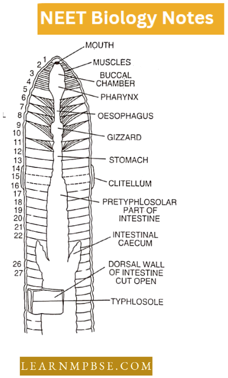
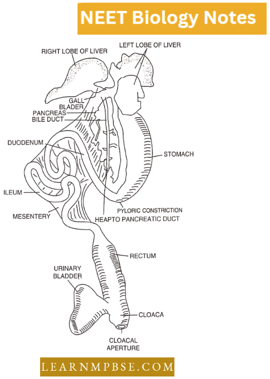
7. Digestive System. The alimentary canal consists of the following parts situated in the segments as mentioned.
- The mouth is situated on the ventral side of the first segment. It opens into the buccal cavity which lies in the first 3 segments. This opens into the pharynx situated in the fourth segment.
- From the 5th to the 7th segment is the oesophagus followed by a highly muscular structure known as gizzard situated in the 8th segment. From the 9th to 14th segments lies the stomach.
- From the 15th segment to the last segment is the intestine. The intestine is divided into 3 parts known as:
- Pretyphlosolar region. (15th to 26th segments)
- Typhlosolar region. (27th to near end except for last 20-25 segments)
- Post-typhlosolar region. (situated in the last 20-25 segments).
8. Excretory System. Excretion takes place through a large number of coiled tubular structures known as nephridia. These are the excretory units. There are 3 types of nephridia found in earthworms:
- Ventral view of earthworm
- Anterior region
- A seta
- Genital area (Ventral view)
- Integumentary Nephridia: These are mesonephric, present on their)her side of the body wall in all the segments except the first two segments. Their number is 200 to 250 in each segment except in clitellum, where they are 10 times more. These arcs are known as “Forests of nephridia.”
- Septal Nephridia: These are enteronephric and situated behind the 15th segment attached to the septa, on both sides. Each row has 80 to 100 septal nephridia. They are enteronephric.
- Pharyngeal Nephridia: They are enteronephric. Three groups of pharyngeal nephridia are situated in each of the 4th, 5th and 6th segments, Out of all the 3 types of nephridia, only septal nephridia have a funnel-shaped structure known as nephrostome and thus can collect the waste products from the coelomic fluid, other two types don’t have nephrostome.
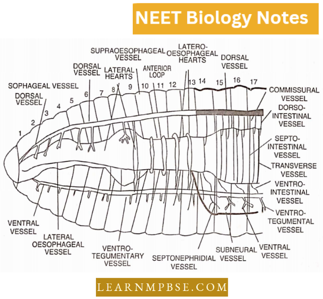
9. Vascular System. It consists of a closed tube of the blood vascular system. The haemoglobin is found dissolved in the plasma. It consists of hearts, blood vessels, anterior loops and blood glands.
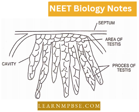
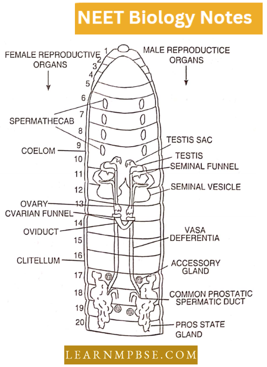
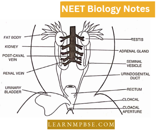
10. Male Reproductive System. Earthworm is a bisexual (hermaphrodite) animal. The male reproductive organs consist of two pairs of testes situated in the 10th and 11th segments.
- Each testis is enclosed in a fluid-filled testis sac. There are 2 pairs of seminal vesicles situated in the 11th and 12th segments. The testes sacs communicate with corresponding seminal vesicles.
- Just beneath each testis lies a funnel known as a spermicidal funnel. From each funnel is given out a duct known as vas deferens. The two pairs of vasa deferentia open into the prostate glands lying from 17th to 20th segments.
- From each prostate gland is given out a duct known as a common prostate spermatic duct. These ducts open on the ventral side of the 18th segment through male genital apertures.
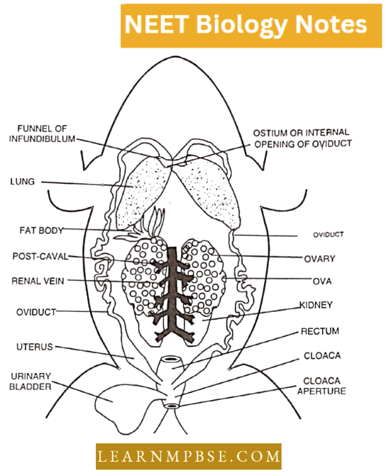
11. Female reproductive organs consist of one pair of ovaries situated in the 13th segment. Just close to each ovary is a funnel, known as the oviducal funnel. Both the oviducal funnels open into oviducts.
- Both the oviducts enter into the 14th segment and open outside through the female genital aperture. Female reproductive organs also include 4 pairs of spermathecae situated in the 6th, 7th, 8th, and 9th segments.
- The spermathecae open to the exterior through 4 pairs of spermathecal pores situated in the inter-segmental grooves of 5/6, 6/7, 7/ 8 and 8/9 segments.
Structural Organisation In Animals For NEET Biology Periplaneta Americana (Cockroach)
Cockroach belongs to the phylum Arthropoda and class Insecta.
Habitat and Habits:
The cockroach is nocturnal, being active at night. Throughout the day, they conceal themselves beneath stones, moist foliage, timber, and various forms of trash.
- Domestic cockroaches inhabit residences, particularly kitchens, storage areas, latrines, sewage systems, ships, and railway cars.
- Certain species inhabit ant nests, while a few are semiaquatic.
- It is highly voracious and omnivorous, consuming any form of animal or vegetable matter that it encounters. It prefers warmth, as it is intolerant of cold. It is a cursorial animal.
The sexes are distinct. It exhibits sexual dimorphism. Life history exhibits steady transformation.
Animal Tissues NEET Notes
Features of cockroach:
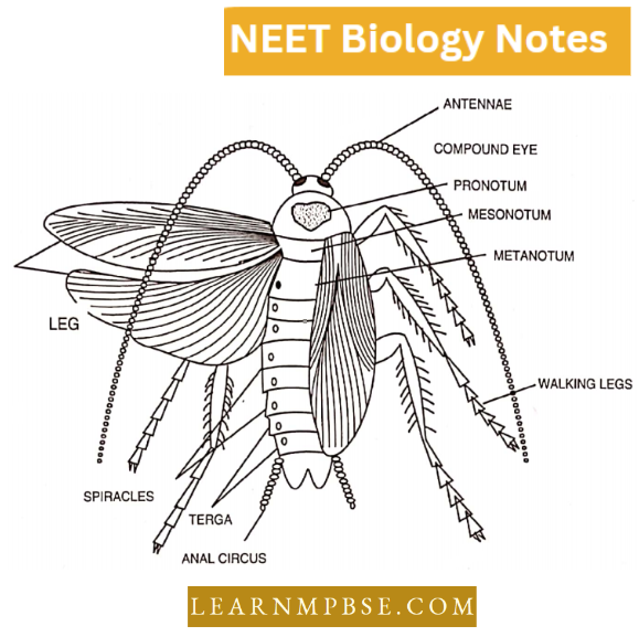
- Shape and size: The body of a cockroach is flat and broad. It is bilaterally symmetrical. The length of the adult is about 1” to 2”, while the breadth is about 1/2″.
- Colour: The colour is reddish brown while there are two dark patches surrounded by a light-brown margin in the first thoracic segment.
- Exoskeleton of Cockroach: The entire body of the cockroach is covered with a thick, hard, dark-coloured chitinous cuticle. It forms the exoskeleton which is secreted by the epidermis.
Thus the body wall is sclerotized or hardened to form sclerites.
These sclerites are joined together by the soft flexible membrane called suture by which movement of the body segment and appendages is possible. Thus, the body is segmented.
- The body of a cockroach is divided into three parts: Head, thorax and abdomen.
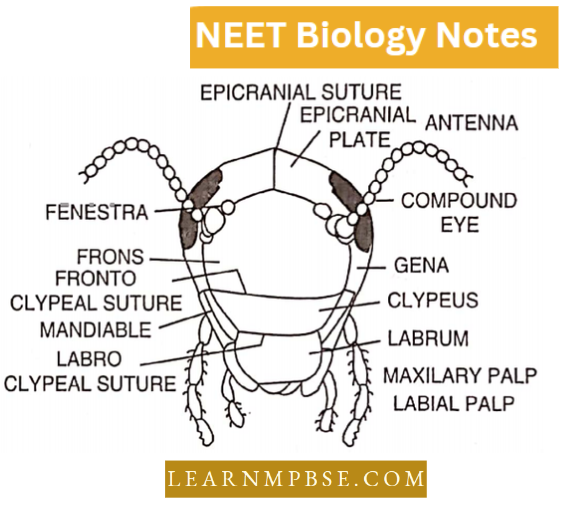
- The head of the cockroach is a hypognathous, strong head capsule or cranium.
- The Head has three types of sense organs: eyes, antennae and fenestrae.
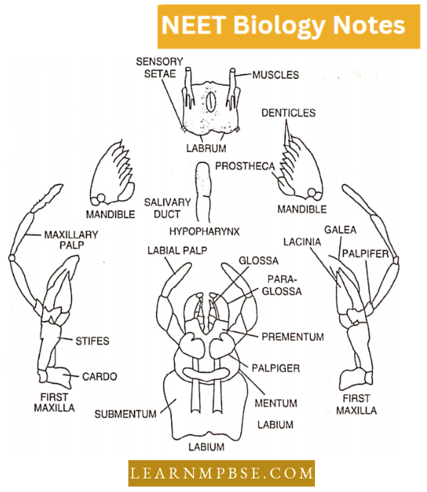
- The head has biting and chewing-type mouth parts which are formed of a labrum, one pair each of mandibles and first maxillae, labium and hypopharynx.
- Mouth-parts: Those parts, which surround the mouth, are called mouth-parts. The cockroaches are as follows :
- Upper lip or labrum.
- Mandible
- First maxillae
- Second maxillae
- Hypophar ynx or lingua.
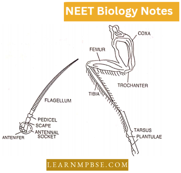
- The Thorax of cockroach bears three pairs of legs and two pairs of wings or pereopods.
Each leg is formed of five podomeres.
1. Wings of Cockroach: There is a pair of wings in the mesothorax and metathorax. These are movably joined to the anterior comers of the terga.
- Each wing consists of membranous expansions of the cuticle thickened by a network of chitinous ridges or nerves or veins which support the wings.
- The anterior mesothoracic wings are called forewings which are dark, opaque, home. Forewings are not used in flight but in a state of rest, they protect the living-wings by overlapping them.
- Hence, these wings are known as fore wings or wing-covers elytra or tegmina. The hindwings or metathoracic wings are thin, membranous, transparent broad, delicate and are used in flight.
- When at rest they are kept folded and laid back over the abdomen under the elytra or wing covers.
2. Leg of Cockroach: There are three pairs of legs, each leg consists of
- Basal podomere or coxa which articulates with the sternum,
- Small trochanter or articular joint which is fused to
- Enlongated and fairly shout femur,
- A slender and spiny tibia and
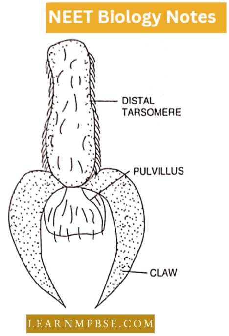
- The tarsus or foot is composed of five short movable podomeres. The last podomere ends in the two claws, also known as pretarsus. Between the two claws, there is a delicate porous pad called the arolium or pulvilus, which is densely covered with bristles and helps in clinging.
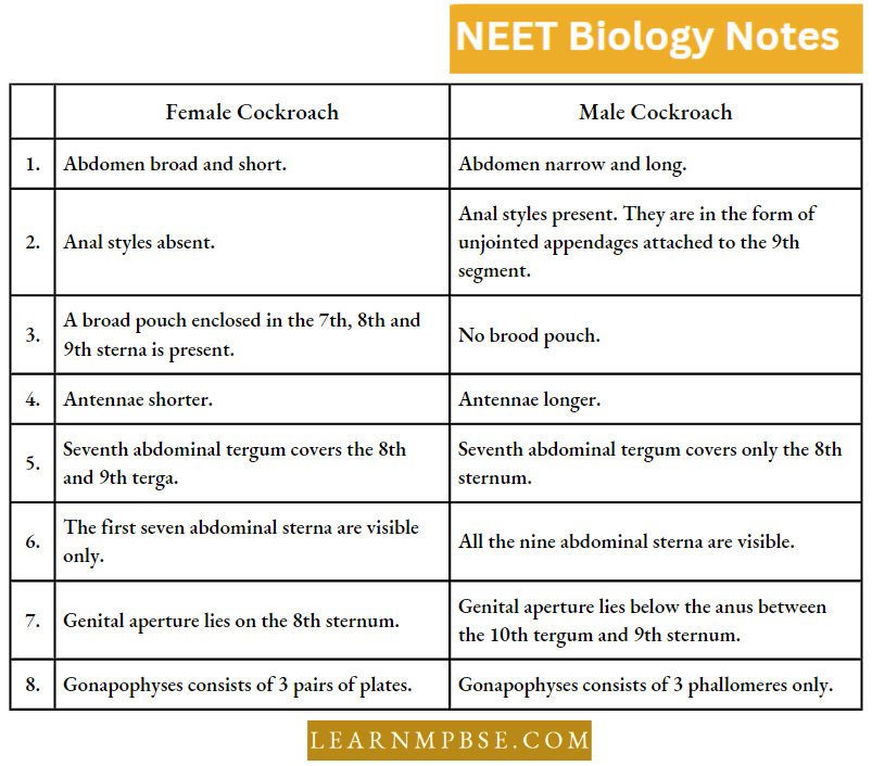
3. The abdomen of cockroaches in both sexes bears one pair of anal cerci and apophyses. A male cockroach has anal styles while a female cockroach has brood pouches.
Structural Organisation In Animals Neet Notes Differences between male and female cockroaches
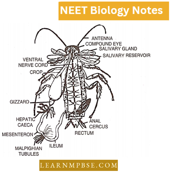
4. The alimentary canal is made up of stomodeum (Foregut). Mesenteron (Midgut) and proctodaeum (Hindgut). The stomodeum and proctodaeum are lined internally by ectoderm whereas the mesenteron is lined by endoderm.
- The foregut consists of the mouth, pharynx oesophagus, crop and gizzard. In the gizzard are present 6 hard cuticular teeth which help in the mastication of the food. There is a stomodaeal valve present in the posterior region of the gizzard.
- The middle part of the alimentary canal is known as the mesenteron. At its anterior end are present 7-8 blindly ending tubular structures known as mesenteric caecae and at the posterior end are present the malpighian tubules.
- The hindgut is made up of the ileum, colon and rectum. One pair of salivary glands are present at the antero-ventral side of the crop.
- Each gland has a reservoir and bipartite glandular portion. A common efferent salivary duct from both glands opens at the base of the hypopharynx.
5. The Circulatory System is of open type. The blood is colourless, without haemoglobin, known as haemolymph. The body cavity (Haemocoel) is divided into 3 cavities by two diaphragms known as pericardial sinus, perivisceral and perineural sinuses.
- The heart is situated in the pericardial sinus and is made up of 13 segmentally arranged funnel-shaped chambers.
- There are fan-shaped muscles attached to the hearts in each segment known as alary muscles which help in opening and closing the ostia. The haemolymph flows in the heart in the forward direction.
6. Excretion is carried out by about 60 fine, yellow, unbranched, thread-like blind tubes called malpighian tubules. The main excretory product is uric acid.
- Respiration is carried out through the trachea.
- Compound eyes are formed of many optical units called ommatidia and the image formed is mosaic.
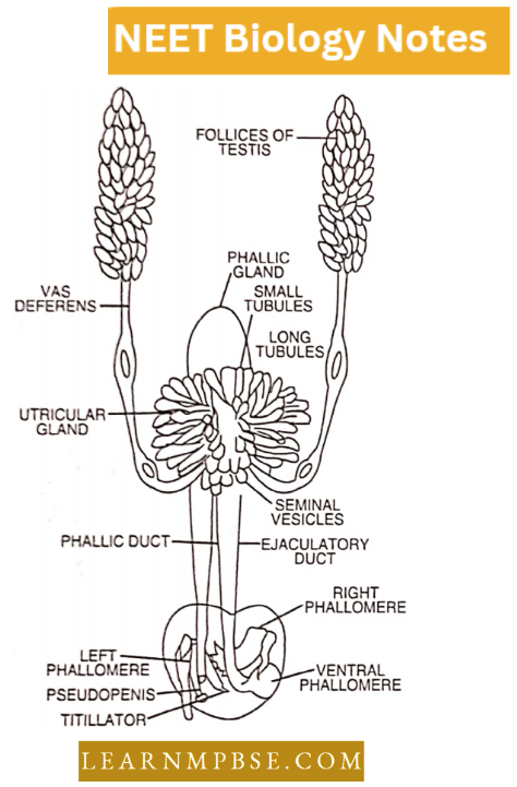
7. Male reproductive organs. It consists of one pair of testes, situated from 3rd to 6th abdominal segments. Each testis gives out a vas deferens. Both the vasa deferentia run posteriorly and open into an ejaculatory duct, where lies a gland known as the utricular gland.
- This gland has 3 types of tubules known as long peripheral tubules, short central tubules and one pair of seminal vesicles which store the sperms. The ejaculatory duct runs posteriorly and opens outside through an aperture known as the male gonopore.
- Around the male gonopore are situated a few chitinous structures known as male gonapophysis. This is made up of 3 phallomeres—left, right and ventral phallomeres.
- The left phallomere has some structures known as Titilator, Pseudopenis and Asperate lobe. All these help during copulation. The sperms of cockroaches are released in the form of a bundle known as spermatophore which is surrounded by 3 layers.
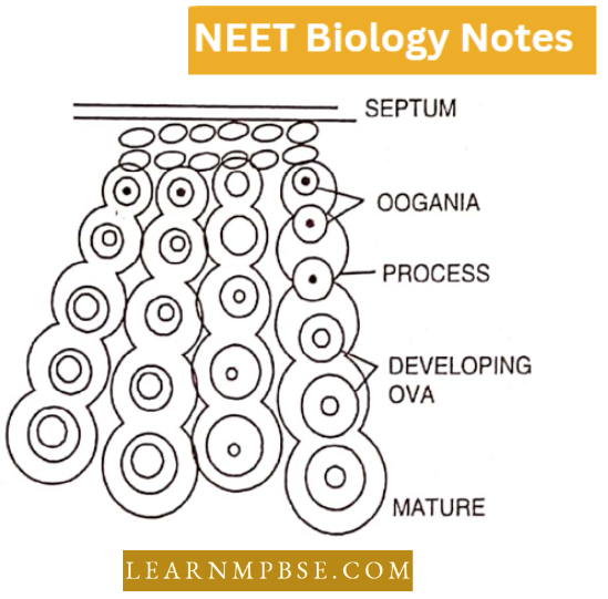
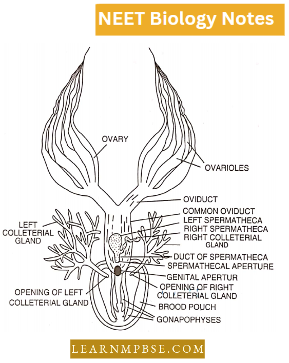
8. Female Reproductive organs. It consists of one pair of ovaries lying from the 3rd to 6th abdominal segments. Each ovary is made up of 8 beaded structures known as ovarioles, in which the eggs are arranged linearly in order of development.
- From each ovary arises a duct known as the oviduct. Both the oviducts join to make a common duct known as the vagina, which opens into the genital chamber through the female gonopore.
- In the gonopore lie a pair of spermathecae, one pair of collateral glands and six chitinous plates known as ovipositors.
9. Ootheca formation and egg laying. The eggs are laid in a brown, home purse-like egg case called an ootheca. The egg cases are laid in a dark place in a crevice, in the dirt, pushed down by the ovipositor.
- Each egg case contains 16 fertilized ova (eggs) in two vertical rows. Each egg in the egg case hatches into a colourless young cockroach called a nymph.
- Nymph, hatched undergoes gradual metamorphosis (6-9 moults) to form an adult.
Structural Organisation In Animals For NEET Biology Rana Tifirina (Frog)
Habitat of frog: Habitat means the natural abode (home) of an animal. Rana tigrina is commonly found in or near water of ponds, lakes, ditches, wells etc. It is an amphibious animal.
Habits
- Feeding: The frog is mainly insectivorous. It feeds on living and moving insects, spiders and worms. It also shows cannibalism as the larger frogs eat up the smaller ones. It captures its prey with the help of its tongue.
- Locomotion: It is through swimming-leaping and floating.
- Croaking. During the rainy season, a noisy chorus of croaking frogs is often heard in the evening or at night. When the air from the lungs is exhaled, it sets the vocal cord, and presents the laryngotracheal chamber, with vibrations and thus voice or croaking is produced.
- The male frogs croak more loudly than the females. They possess a pair of large, distensible vocal sacs, one on either side of the throat. The vocal sacs act as resonators and help in raising the mating call for the female.
- Breeding: During the rainy season (i.e. from July to September) frogs collect in some quiet, shallow water pond and croak loudly. The male and female frogs undergo pairing.
- The swellings called copulatory or nuptial pads help in clasping the female firmly. The couple remains in this condition for several days and can swim in water; the female mainly bears the burden of the male during swimming.
- The female ultimately lays an egg over which the male sheds its spermatic fluid. The sexual embrace between a male and a female frog which leads to the laying of the egg by the female and the discharge of sperm by the male is called amplexus or pseudocopulation.
- It differs from the process of copulation in which the male transfers its sperm inside the body of the female. After amplexus, the male frog gets down the female and both start living separately.
- Hibernation. A frog is a cold-blooded or poikilothermal animal. Its body temperature does not remain constant, but it changes according to the temperature of the surroundings in which it lives.
- In winter, the temperature of its body falls considerably. It cannot bear the severe cold of winter. Its metabolic activities are slowed down and this may ultimately lead to death.
- To avoid this, it buries inside the mud or moist soil. It may burrow about 60-90 ems deep. There it lies in a state of rest or sleep. It does not move. Its mouth and nostrils are kept closed.
- It respires through the skin. It does not feed and lives on food stored as glycogen in the liver and fat in the fat bodies. In this condition, it rests or sleeps throughout winter.
- This particular condition of rest in winter by frogs is called winter sleep or Hibernation. At the approach of spring, warmth wakes it up and it comes out of its burrow and again begins to lead an active life.
- Aestivation. During summer, when the temperature is quite high, the frog again goes deep into the water. It may bury itself in the mud at the bottom of the pond. The pond may dry up. The frog remains inactive and can tide over the unfavourable period.
Morphology And Anatomy Of Animals NEET
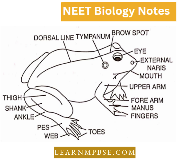
Structural Organisation In Animals Neet Important Questions Size, Shape and Colour
- Frog has an ovoid and dorsoventrally flattened body which is streamlined. It has a length of 10-17 cm and a breadth of 5-8cm.
- The body shows external bilateral symmetry whereby the right and left halves of the body are mirror images of each other. The animal has anterior, posterior, ventral, dorsal and lateral sides.
Body Covering
- The body is covered over by a loosely attached skin. The surface of the skin is smooth and soft. It is also slippery because it secretes a thin film of mucus.
- Due to the looseness of the skin, several longitudinal wrinkles appear especially on the dorsolateral sides. They are called dermal plicae.
Body Divisions
- The body of the animal can be divided into two parts, head and trunk. The tail and neck are absent.
- To the anterior and posterior parts of the trunk are attached limbs, one on either side.
Head
- The head of the frog is roughly triangular in outline. It is flattened. The anterior upper part of the head is called the snout.
- The snout is blunt. The mouth is in the form of a transverse slit which runs throughout the border of the head.
- It is normally kept closed except at the time of the feeding. Just behind the tip of the snout are found two small dorsal openings called external nares or nostrils. The top of the head bears two dorsolateral large and bulging eyes.
The position of the eyes enables frogs to see in different directions even in the absence of a neck. Each eye has three eyelids. – upper, lower and nictitating membrane.
- The upper eyelid is opaque, pigmented and thick. It is almost immovable. The lower eyelid is small and semitransparent. It is slightly movable. The nictitating membrane is also called the third eyelid.
- It arises from the inner surface of the lower eyelid. The nictitating membrane is transparent and freely movable to cover the whole eyeball. It protects the eyeball from mud and water.
- Outside water, keeps the eyeball moist. The eye can be withdrawn into the head. Just anterior to the eyes, is found a small dorsal and light-coloured swelling called a brow spot.
Behind each eye is found an obliquely placed circular membranous patch named tympanum ear drum or tympanic membrane. It is bounded using a slightly raised ring of cartilage. External pinna or ear is absent.
- The ventral surface of the head is soft and is called the throat. It is alternately raised and lowered in a type of breathing.
- In males, the throat beats two bluish and wrinkled particles towards the posterior end. They are called vocal sacs. The vocal sacs can be Inflated to act as resonators of sound.
Trunk
- The main body or trunk is flattened and oval. It can be distinguishable Into the anterior hard chest and posterior sort belly or abdomen.
- The posterior end beats a small circular apci line called the cloacal aperture. It is a common opening for the passage of animal excreta, urine, eggs or sperm.
Pairs of Pcntadactyl Limbs
The trunk bears two pairs of appendages, forelimbs and hind limits. Therefore, Hie’s animal is a quadruped. The lore limb has three parts-upper arm or hrucliltim, fore units or antebradiium and hand or maims.
- The hand is further distinguishable into three parts carpus, palm or metacarpus and fore-diglsts or lingers. There are only four fingers. The finger corresponding to our thumb is absent.
- However, it can fall under the skin. The first or inner among the remaining four is equivalent to our index linger.
- This finger develops ampleuxsory or nuptial pad during the breeding season in males, Articular pads occur on the under-surface of the finger joints.
In the silling position die lore limbs arc bent outwardly in the elbow region. The hind limbs or legs are differentiated into a proximal thigh or femur, a middle shank or crus and a distal fool or pes.
- The foot has diree parts ankle or tarsus, metatarsus or instep and live toes or bind digits. The membranous fold of skin or web occurs between die toes. The first toe is called hallux.
- The fourth toe is the longest. A prehnlhix or sixth toe can fall under the skin, Articular pads occur on die under-surface of toe joints. In the selling position, the hind limbs are folded like the letter Z.
Rattus Rattus (Rat)
- Common Indian black rat (Rattus rattus) in nocturnal, inhabiting holes and burrows.
- A rat is about 20 cm in length, its body is covered by hair, except in a few places and is characterized by a fusiform body.
- The rat is bilaterally symmetrical. Its body is covered over by soft fur of hairs shown as pelagae. It is black dorsally and grey on its ventral side.
Features of Rat
- Its body is 15 to 20 cm in length and is divided into 4 regions.
- Head
- Neck
- Trunk
- Tail
- The integument is made up of epidermis, dermis and their derivatives.
Rat External characters.
1. Head. It lies at an angle with the long axis of the rest of the body. It is elongated and conical. It is broad posteriorly and tapers anteriorly into a naked snout.
- At the tip of the snout, is a short, transverse opening, known as the mouth, The mouth is bounded by fleshy and movable upper and lower lip. The upper lip bears a cleft in the middle to form the hair lip.
- Through this cleft of the upper lip, the incisors are visible. Just above the mouth, are situated two inverted comma-like nostrils or external nares, which lead into the nasal chambers.
- Behind the nostrils, on the side of the head, lies a prominent bulging eye. The eyes are black and the pupils are rounded. Each eye is bounded above, and below by movable and opaque upper and lower eyelids.
The eyelids are provided with very fine and short eyelashes. The third eyelid i.e., the nictitating membrane is also present at the inner corner of each eye.
- The meibomian glands, which are situated at the margin of the eyelids, are very prominent or conspicuous. Behind the eyes, the head bears posterolaterally, a pair of external cars or pinnae. The pinnae are meant for collecting sound waves.
- On the sides of the snout and also above the below eyes, are present long, stiff, bristle-like hairs, called whiskers or vibrissae.
- They are sensitive to touch and they enable the animal to judge whether it can pass through a
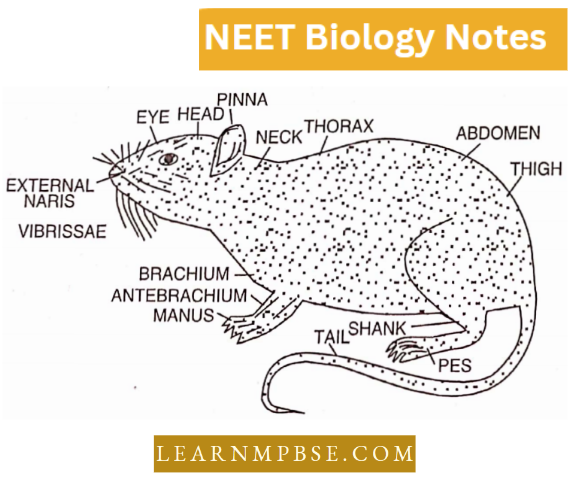
2. Neck. It is short and without anything special. It simply connects the head with the trunk and enables the former to move in all directions.
3. Trunk. It is lightly compressed dorsoventrally and divisible into two regions.
- Thoracic region.
- It is the anterior small region of the trunk and is also known as the chest.
- It is strengthened by the ribs.
- It encloses the heart and the lungs.
- Abdominal region.
- It is the posterior larger and soft region of the trunk.
- It is without ribs and encloses the remaining viscera of the body.
- On the ventral side of the trank of females, are present six pairs of teats or nipples or mammae on which open the milk glands.
Three pairs of teats are present in the thoracic region and the remaining three pairs are restricted to the abdominal region of the trank. In males, the teats are vestigial.
There are two pairs of limbs, attached at the anterior and posterior sides of the trank. The limbs are constructed on the pentadactyl pattern.
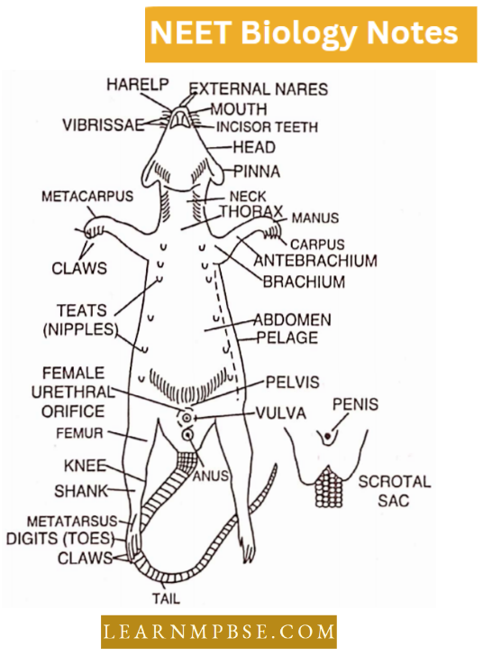
Each forelimb consists of an upper arm or brachium, forearm or antebrachial and hand or manus. The hand has a wrist or carpus, palm or metacarpus and 4 well-developed lingers or digits.
- The thumb is vestigial. Each digit ends in a horny claw, which is well developed and is used for digging the burrows.
- Each hind limb is divided into a thigh or femur, a shank or crus and a foot or pes. The foot consists of an ankle or tarsus, insteps or sole or metatarsus and 5 clawed digits to toes.
- The typical walking pads are present on the tips of the digits, palms and the insteps. The palms and soles are not hairy.
At the posterior extremity of the trunk, underneath the base of the tail, lies an opening, the anus. It is the posterior opening of the alimentary canal.
- In front of the anus, oh the ventral side of the trunk, are present the external sex organs. In male rats, they consist of a pair of scrotal sacs, containing testes and a retractile penis, the copulatory organ.
- The distal part of the penis is very sensitive and known as the glans penis, covered over by a loose fold of integument, the prepuce. At the tip of the penis is situated the urinogenital opening.
- In female rats, the sex organs are marked by an opening of the vulva which leads into the vegina.
In front of the vulva, is present the urethral orifice, situated on a short papilla. Unlike male rats, the urinary and genital openings are separate in female rats.
4. Tail. It is long and covered with small overlapping scales. Emerging from under the leg of each scale, are three short bristle-like hairs. The tail is a balancing organ.
Structural Organisation In Animals Neet Previous Year Questions Interaction of Earthworms with mankind
The Earthworms are both beneficial and harmful to man directly or indirectly.
Beneficial aspects: The beneficial aspects of the Earthworm are as follows:
1. Beneficial to agriculture: The Earthworms are of enormous value to agriculturists in the following ways:
- The burrows of the Earthworms make the soil more combed which becomes porous and serves better for respiration and penetration of the developing roots. The earthworms are considered to be natural ploughs.
- The decaying vegetation which is carried by Earthworms into the burrows enriches the soil.
- The soil particles are broken into a fine state when they pass through the muscular gizzard.
- The Earthworms bring out fresh soil in the form of castings and thus a thick layer of humus is formed. Along with the castings, nitrogenous matters are also given out which are added to the soil and used by the plants.
- According to Charles Darwin, there are about 53,000 Earthworms in one acre of soil.
- He estimated that in a single year, about 80 tons of castings are added to the surface of soil in one acre and can cover the surface by a layer of about 1/5 inch. Thus the earthworms are known as friends of the farmers.
2. As food for man: In many countries such as China, Japan, Burma etc. Earthworms are used as food for man. The edible preparations are made in many ways.
3. As medicines: Earthworms are also used as medicines. It is said that it cures stones in the bladder, jaundice, piles, diarrhoea, sexual impotency etc.
4. As bait for fishing: Earthworms are also used as bait for fishing. These are cut into small pieces and used as baits. In many countries, there are separate centres for the sale of earthworms.
5. Use in the laboratory: As earthworms are commonly found all over the world they are used for dissections in zoological laboratories.
6. As a dispersal agent: The dispersal of small seeds and fruits is also done by Earthworms.
Harmful aspects: The Earthworms are also harmful in the following ways:
- They spoil the fields and the gardens. Burrows often cause water-oozing and soil erosion.
- Earthworms also act as intermediate hosts of some parasites such as Monocystis, the cestoda, and Amoeba. Taenia and the Metastrongilus.
- The Earthworms also damage the plants. Pheretima damages the roots of Piper Betel in Coimbatore. It also damages the roots of paddy in Malabar. However, the Earthworms are more beneficial to man.
Structural Organisation In Animals Neet Mcqs With Answers Interaction of Cockroach with Mankind (Economic Importance)
- Harmful activities
- It damages household materials like clothes, shoes, purses, etc.
- It contaminates the foodstuffs and befouls them so that they become unpalatable.
- These also act as vectors for the germs of some diseases like cholera, diarrhoea, typhoid, etc.
- Useful activities
- These are used as food by the people of some South American countries and Myanmar.
- These are used as food for many useful animal types like amphibians, lizards, birds and rodents.
- Cockroaches are used as safe experimental animals for laboratory experiments and biological research as these are easily available.
Structural Organisation In Animals Neet Revision Notes Interaction of frog with mankind (Economic Importance)
A frog is a useful animal for to following reasons:
- A frog is used in biological control as eats upon crop’s pest insects which also saves the expenditure on the insecticides.
- A frog is used as an experimental animal for teaching, research, pharmacology, etc.
- Its muscular legs are used as food in some parts of India and many other countries.
- Small froglets are used as fish bait.
Structural Organisation In Animals Neet Study Material Interaction of Rats with Mankind
Beneficial activities:
- It serves as a crucial element of the food chain, providing sustenance for many reptiles, birds, and mammals.
- It is utilized as sustenance for individuals in certain nations.
- It is beneficial in education and biomedical research.
- These are utilized as experimental subjects to investigate the effects of pharmaceuticals for eventual application in human medicine.
- These are utilized in genetic studies to examine traits such as variances in coat color.
Detrimental actions:
- These serve as significant pests of crops and stored grains, inflicting harm on approximately 20 percent annually.
- These entities undermine the roots of plants by creating burrows and tunnels.
- Burrows constructed by rats provide as refuge for venomous creatures. For instance, serpents.
- These inflict harm on books, clothing, food, leather items, doors, and other materials due to their nibbling behavior.
- It serves as a vector host for rat fleas that disseminate the plague bacterium.