
NEET Biology Kingdom Monera Main features
It encompasses all prokaryotic species, with size ranges of 1-10 µm, including cyanobacteria (blue-green algae) and bacteria.
- Prokaryotic cells are distinguished from eukaryotic cells by possessing a cell wall composed of complex substances (excluding cellulose and chitin), circular bare DNA, and the absence of membrane-bound organelles such as mitochondria.
- Some prokaryotes possess internal membranes, such as lysosomes in certain bacteria, and membrane systems that house enzymes for photosynthesis and respiration.
- Prokaryotes reproduce via binary fission.
- Cyanobacteria do not exhibit sexual reproduction; rather, genetic recombination in certain bacteria transpires through conjugation, transformation, and transduction.
- Numerous prokaryotes produce spores in response to unfavorable conditions.
Read and Learn More NEET Biology Notes
- Their small size, metabolic diversity, and rapid reproduction probably account for the evolutionary success of prokaryotes.
- Classification of Kingdom Monera. Simple prokaryotic cell structure, unicellular, both heterotrophs and autotrophs. Kingdom Monera was created by Copeland (1966), and divided into three main groups.
- Schizomycophyta (Bacteria) smallest living cells, basically unicellular, with indistinct nuclei, colorless, and mainly heterotrophic Examples; are Cocci, Vibrio, Bacilli, Spirilla.
- Cyanophyta or Cyanobacteria (Blue-green algae). Unicellular, no distinct nucleus, blue-green color e.g. Oscillatoria, Nostoc, Gloeoccipsa, Tolypothrix.
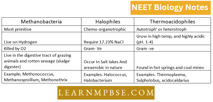
- Archaebacteria. Most primitive prokaryotes are considered to evolve immediately after the evolution of first life, The cell wall lacks peptidoglycan but contains protein and non-cellulosic polysaccharides. Branched-chain lipids in membrane. Live under hostile conditions Example; Methanogens, Halophiles, and Thermoacidophiles.
Kingdom Monera NEET Notes
NEET Biology Kingdom Monera Bacteria
- Bacteria are very small and their size generally ranges from 0.2 – 1.5 pm in diameter and 2 – 10, pm in length.
- Dialister pneumonsinilis is the smallest bacterium (0.15 — 0. 3 pm long); Bacillus buischili (80 pm long, 3 — 6 pm, diameter); Beggiatoa mirabilis is the largest bacterium (16 — 45pm diameter and upto several centimetres long).
- Bacteria occur in three basic forms or shapes. These are either spherical (cocci), rod-shaped (bacilli), or spiral (vibrio/spirillum). Though most bacterial species have cells that are of a fairly constant and characteristic shape, some species are pleomorphic (i.e.; these can exhibit a variety of shapes).
- Gram staining, introduced by Christian Gram in 1884, divides bacteria into two groups: Gram-positive and Gram-negative.
- Flagella (sing, flagellum; Latin whip). Long (3 — 12 pm), fine, wavy, filamentous appendages that protrude through the cell wall, responsible for the motility of bacteria.
- These are much thinner than the flagella or cilia of eukaryotes, being 0.01 to 0.02 pm in diameter.
- Bacteria may be atrichous (without flagella), monotrichous (with one flagellum), lophotrichous (with a group of flagella at one end), amphitrichous.
- Two flagella (also two groups of Flagella), one at each end Example; Alkaligenes feces and peritrichous (flagella present all over the body) ccphalotrichous—(Two or more flagella at one end)
- Pili or Fimbriae (Sings. Pilus = hair fimbirae = fringe). These are hollow, nonhelical, filamentous appendages projecting from the walls of some gram-negative bacteria.
- These are thinner and shorter; and more numerous than the flagella. These also arise from the basal body and are made up of specific proteins called pilin.
- Some bacterial cells are surrounded by a viscous substance forming a covering layer or envelope around the cell wall. If this layer is in the form of a loose mass, it is called slime.
- Below the external structures like capsules and flagella; and outside the cell membrane is present a rigid structure called a cell wall. Due to its rigidity, it protects the internal structures of the cell and provides shape to the cell.
- However, its main function is to prevent the cell from expanding and bursting (Most bacteria live in hypotonic environments, and are likely to take in much water and eventually burst).
- The cell walls of almost all the eubacteria (true bacteria) are made up of peptidoglycan, also called murein or mucopeptidc. It is found only in monerans.
- The plasma membrane is a phospholipid membrane also containing proteins and polysaccharides. It is selectively permeable. The respiratory enzymes are associated with the cell membrane as the mitochondria are absent.
- Mesosomes. The cytoplasmic membrane is invaginated at certain places into the cytoplasm in the form of the system of convoluted tubules and vesicles, (mesosomes).
- On their surface are found enzymes associated with respiration. Therefore, these are supposed to be analogous to the mitochondria of eukaryotes.
- The cytoplasm is a complex mixture of amino acids, proteins, lipo-complexes, nucleotides, carbohydrates, vitamins, and coenzymes.
- The membrane-bounded organelles like endoplasmic reticulum, mitochondria, and Golgi bodies are absent in bacteria. However, ribosomes are present in sizeable numbers, providing a granular appearance to the cytoplasm.
- The cytoplasm contains about 10,000—30,000 ribosomes of the 70 S type. The sedimentation coefficient of bacterial ribosomes is 70S. Each ribosome consists of 2 unequal subunits (30S and 50S). Each subunit is composed of RNA and protein.
- The most common non-living inclusions in the cytoplasm are —volutin granules, PHB, and elemental sulfur. The volutin granules, also known as metachromatic granules, are polymetaphosphates and serve as a reserve source of phosphate.
- PHB. (Poly (3 hydroxybutyrate) is a lipid-like material and can serve as a reserve carbon and energy source.
- Some bacteria that live in aquatic habitats form gas vacuoles which provide buoyancy to the cells.
- As compared to the eukaryotes, bacteria do not contain a distinct membrane-bounded nucleus and all other membrane-bound organelles are absent.
- However, an amorphous lobular mass of fibrillar, chromatin-type material that occupies about 10 — 20% of the cell, is present near the center of the cell.
- DNA of the cell is confined to this area called nucleoid or chromatin body or nuclear equivalent or genophore.
- It is also called a bacterial chromosome as it consists of a single, circular DNA molecule in which all the genes are linked.
- This single molecule is over a thousand times longer than the cell itself and is, therefore, highly folded. Bacteria generally lack the histone proteins.
- In addition to the normal DNA chromosome, some extrachromosomal genetic elements are often found in bacteria. These elements are called plasmids.
- These are circular pieces of DNA that have extra genes. These are capable of autonomous replication in the cytoplasm of the bacterial cell.
- Plasmids carry genes responsible for different functions in bacteria. For example, Rfactor plasmids (which confer resistance to antibiotics), colicincrgic factors (responsible for the formation, in coli. of special proteins that kill closely related species of bacteria) of genes (for nitrogen fixation).
Reproduction
It takes place by binary fission with the cell dividing at right angles to its long axis. The following steps are involved:
- Division of nuclear material. After the bacterial cell has attained maximum size, the nuclear material duplicates and divides without any spindle formation. The DNA is found attached to the mesosomes near the septum-forming region.
- Cleavage of the cytoplasm. The cell membrane from the middle part of the cell grows inwards dividing the cytoplasm into two halves.
- Formation of cross wall. In between the two newly formed cytoplasmic membranes, a cell wall (septum) is laid down, forming two daughter cells.
- Separation of daughter cells. The growth of the two daughter cells creates tension that separates the two newly formed cells.
Kingdom Monera Notes For Neet
Transfer of genetic material (Genetic recombination)
- Conjugation. Investigations with an electron microscope have shown that two E. coli cells can conjugate and the genetic material (DNA) from one cell can be transferred to the other. The sex difference between the Z (donor) and Z (Recipient) cells is determined by the presence or absence of a specific genetic factor called F (fertility factor. When present, F is a donor cell, and when absent, F-, is a recipient cell. The F factor can exist in two alternative states—as part of the E. coli chromosome or as a very small free DNA strand.
- The £. coli strains are then (high-frequency of recombination) or metafile and F strains respectively.
- Conjugation occurs between an Hfr and a cell or between a P and a Pcell. The P cells transfer only the F factors after its replication.
- The cells transfer both the F factor as well as the genetic constitution of the donor cell. The recipient cell now becomes a merozygote (partially diploid).
- Transformation. A genetic change takes place in the recipient bacterial cell as a result of the absorption of DNA fragments reduced
- Transduction. It is a process in which fragments of UNA are carried by bacteriophages train one bacterial cell to another. It was first observed by Zander and Lederherg (1952).
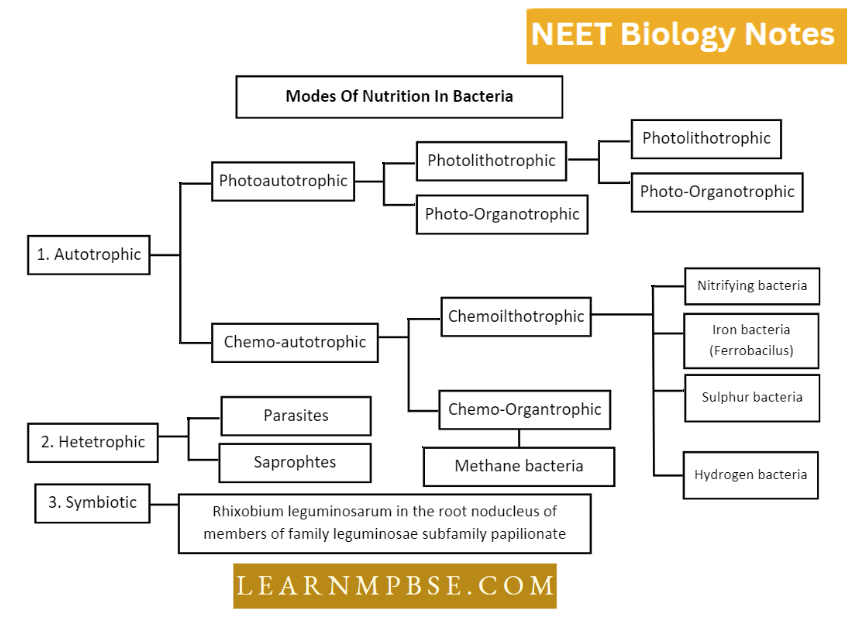
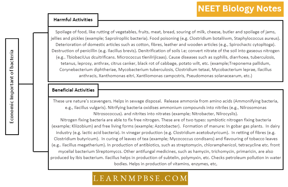
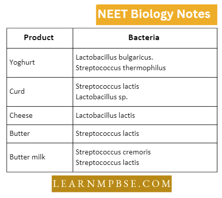
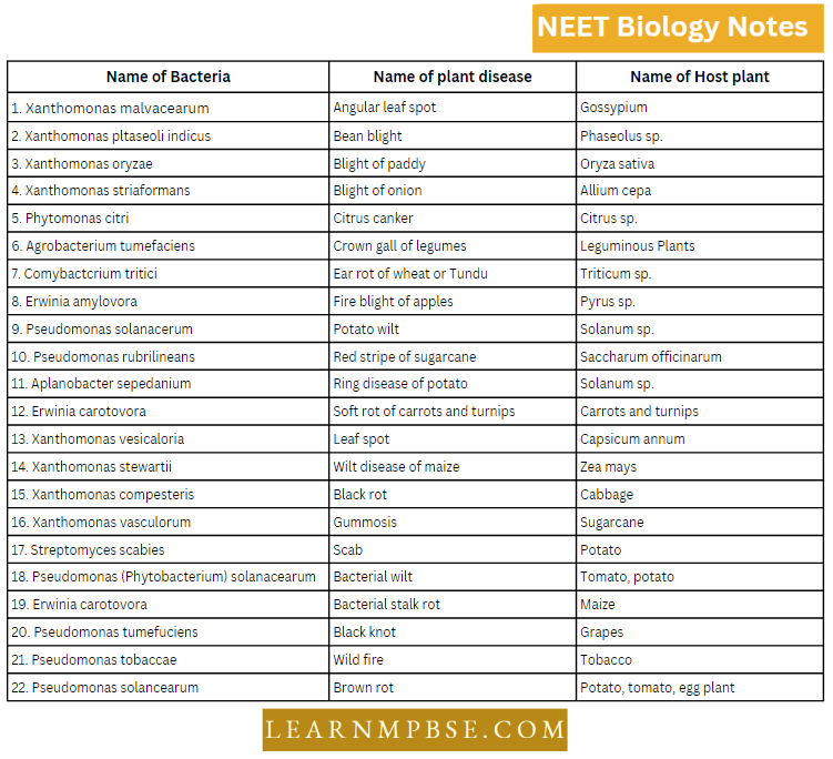
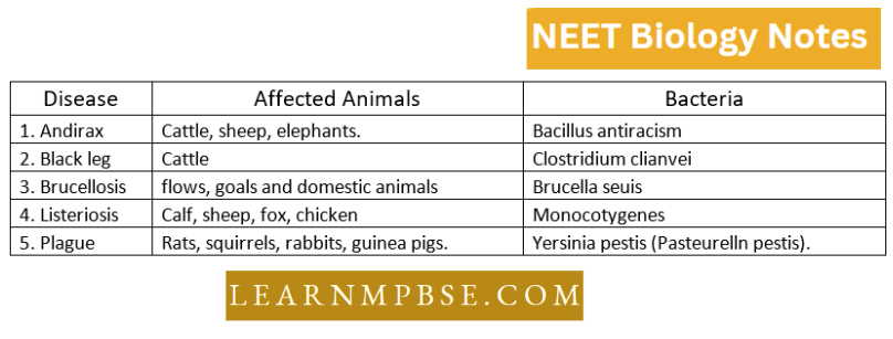
NEET Biology Kingdom Monera Economic Importance Of Bacteria
Cyanobacteria Characteristics NEET
NEET Biology Kingdom Monera Actinomycetes
These microorganisms connect bacteria with fungus. Similar to mushrooms, their somatic architecture comprises multicellular filaments (hyphae) that collectively constitute the mycelium.
- They were previously referred to as “Ray Fungi.” They generate asexual spores, referred to as conidia, on aerial hyphae, which serve as asexual spores in fungi, in contrast to bacterial endospores.
- They are thermally inactivated and not thermally resistant. Their spores and cells do not exceed 2 to 3 microns in diameter.
- Actinomycetes are either saprophytes or parasites. Certain organisms, such as Streptomyces spp., thrive in the soil.
- Conversely, several species, such as Micromonospora spp., are only located in semi-aquatic environments, specifically among lacustrine muds.
- Certain members, like as Nocardici spp., are recognized for their ability to breakdown substrates including paraffin, phenol, and petroleum-derived compounds, which are very resistant to most bacteria.
- Actinomycetes spp. are specialist parasites located in the oral cavities of animals. Reproduction through fragmentation or conidia.
Kingdom Monera Notes For Neet
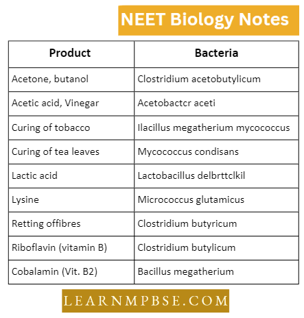
Economic Importance
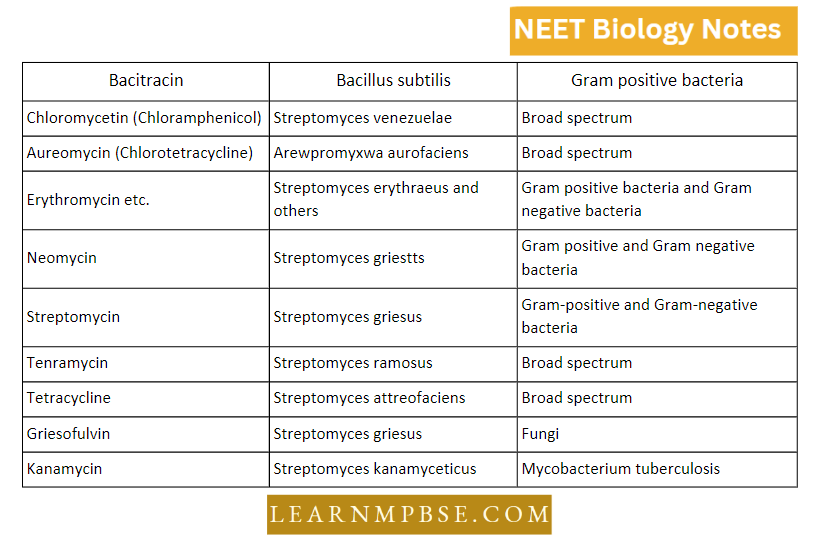
The genus streptomyces of this group is most important, some species of which produce antibiotic substances of great medicinal value like streptomycin, chlorotetracycline, oxytetracycline, tetracycline, chloramphenicol, and erythromycin.
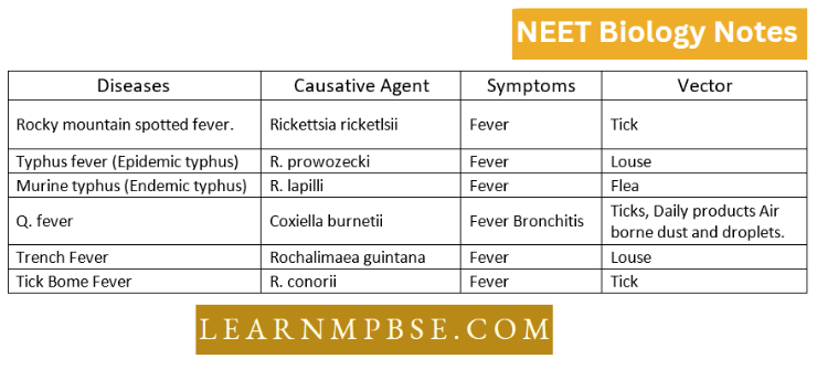
NEET Biology Kingdom Monera Chlamydiae
These are the smallest recognized bacteria and are subgroups of rickettsiae which cause diseases in humans and animals. They have DNA, RNA, and some enzymes. They can be treated with some antibiotics.
The two species of chlamydiae are:
Chlamydia trachomatis causes trachoma in the eyes and two sexually transmitted diseases such as chlamydial urethritis and lymphogranuloma encrust.
Chlamydia psittaci which causes psittacosis in birds. L-forms. Klieneberger discovered L-forms during the culture of Streptobacillus moniliformis.
being the initial of the Lister Institute named for Lord Lister (a pioneer in aseptic surgery). These micro-organisms lack cell walls and are produced only by Bacillus.
L-forms can be converted again into Strcp-tobacilliis. L-forms can be selected by cultivation of bacteria in an osmotically buffered penicillin-containing medium.
Therefore, L-forms arc bacteria in which the primer for peptidoglycan has either been eliminated or modified by penicillin treatment.
They are found in water and mud as free-living organisms. They form a small group of heterotrophic bacteria. Most of the spirochaetes are parasites of man and animals.
They are unicellular and helicoid in shape. The cell has two overlapping sets of fibrils.
The protoplasm is enclosed by a flexible cell wall.
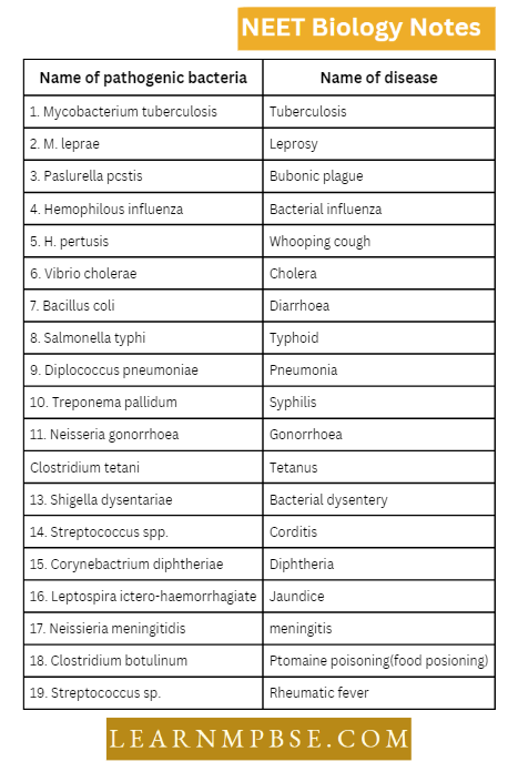
Spirochaetes cause some human diseases as Syphilis is caused by Treponema pallidum Relapsing fever by Borrelia Jaundice (infectious) by Leptospira
NEET Biology Kingdom Monera Archaebacteria (Ancient Bacteria)
They are highly primitive and inhabit exceedingly inhospitable environments where few other species can endure.
- Certain organisms are anaerobic and generate methane, while others inhabit highly saline environments or thrive in hot, acidic sulfur springs.
- Their cell wall lacks peptidoglycan.
- They have special branched-chain lipids in their cell walls which make them heat and acid resistant.
Differences between cyanobacteria, halophiles, and thermoacidophiles
Gram-Positive vs Gram-Negative Bacteria NEET
Differences between Gran +ve and Gram -ve bacteria

Neet Biology Kingdom Monera
- Mirco-organisms like bacteria sometimes can exist without a cell wall. The hu-cell membrane and its intact contents are then called protoplasts (osmotically fragile). Young actively growing gram (+) bacteria are sensitive to penicillin. So. these bacteria can Be made of protoplasts.
- The nrehacbactcria and bacteria possibly arose from a more ancient form of life called progenitor. Understanding one kind of organism requires the isolation of one individual and multiplying or culturing it. i.e. to obtain pure culture.
- A pure culture contains multiple copies of a single kind of micro-organism. The microbiologist has developed several techniques to obtain the pure culture of microorganisms.
- All eubacteria have a cell wall, made up ofmurein or peptidoglycan. It consists of polysaccharides cross-linked with short amino acid chains. Bacterial conjugation though different from eukaryotic sexual reproduction is a means of making new genetic combinations that are expressed as progeny.
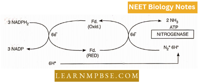
- Rhizobium is a symbiotic nitrogen-fixing bacterium. The non-symbiotic free living nitrogen-fixing bacteria are Azotobacter, Beijerinckia, and Klebsiella (all aerobic) The non-symbiotic free living anaerobe, nitrogen-fixing bacterium is Clostridium pasteurianitm.
- Chromalium. Rhodospirillum arc photosynthetic nitrogen-fixing bacteria. E. coli bacteria are present in the large intestine of humans and mammals. They are straight rods. There are motile and non-motile types of E.coli.
- The cell surface has pili on which certain phages are absorbed. E. coli is a facultative anaerobe. The optimum temperature for its growth is 30-37 °C and the optimum pH value of medium is 7.2— ca> 7.5.
- Nosloc sp. occurs within the tlialli of Blasia and Anthoceros (The Bryophytes). Nostoc sp. lives within the cells of Gcosiphon pyreforme (a fungus) Nostoc sp. occurs in the petiole of Gunnera (an angiosperm). Calothrix sp. lives within the cells of Enteromorpha (a green alga).
- Trifolium alexandrinum (Clover) contains Nostoc in its nodules. The reddish color of the Red Sea is due to a cyanobacterium Trichodesmium erythraeum.
- Cyanobacteria associated with Protista. Death factors VFDF (very fast). FDF (fast) and SDF (slow) are toxins produced by cyanobacteria. Biological nitrogen fixation has been discovered by Winogradsky.
- The smallest cocci range from 0.5 pm to 1.5. pm in diameter while spiriJJi are as large as 60pm (Peberdy, 1980). About one trillion (1 012) bacteria of average size could be packed into a 1 ml pipette.
- A single drop of water may contain as many as 50 thousand million bacteria. A teaspoonful (5 ml.) of packed bacteria represents 2000 times as many individuals as there are people on Earth.
- A tea .spoon of rich soil contains billions of bacteria Mycobacterium and Xanthomonas form nodules in the leaves of Ardisia and Puvetta while Frankia forms nodules in the roots of Alms and Casuarina.
- Gram-negative bacteria are usually partly or wholly resistant to penicillin.
- When Gram-positive bacteria are treated with lysozyme (found in egg white, secretion of skin and mucous membranes and tears) they are rapidly denuded of their cell walls and become naked protoplasts while the peptidoglycan cell wall of Gram-negative bacteria is protected by an outer layer of lipo-complex (it can be removed by ethylene diamine tetra acetate or EDTA).
- So the cell wall of Gram (—) bacteria is not completely re¬ moved. Such only partially denuded cells are called spheroplasts.
- The cell walls of Gram-positive and Gram-negative bacteria differ in their chemical composition. In Gram-positive bacteria, the cell wall has a thick peptidoglycan layer that comprises 90 percent of the cell wall.
- The cell walls of gram-negative bacteria are much more complex. The peptidoglycan layer is very thin making up only 10% or less of the cell wall.
- However, the most interesting feature is the presence of an outer membrane that covers a thin underlying layer of peptidoglycan. The outer membrane is a bi-layered structure consisting chiefly of phospholipids, proteins, and lipopolysaccharides (LPS).
- Due to the presence of the outer membrane, Gram nega¬ tive bacteria are rich in lipids that make up about 11-12% of the dry weight of the wall. Teichoic acid is absent.
- The cells of certain bacteria like Aquaspirillum magnetotacticum contain structures composed of iron in the form of magnetite (Fe304). These are called magnetosomes and help bacteria orient themselves along the geomagnetic lines.