
NEET Biology Animal Nutrition And Digestive System
Nutrition: Nutrition is the procurement of substances termed nutrients necessary for growth, maintenance and providing energy to carry out synthetic activities of living bodies.
Nutrition is of two types :
- Autotrophic mode of nutrition.
- Heterotrophic mode of nutrition.
Autotrophic Nutrition. Mostly green plants can manufacture their organic food due to the presence of chlorophyll. They take up CO2 and H2O and manufacture carbohydrates in the presence of sunlight.
- Such organisms are called autotrophs and their mode of nutrition is called autotrophic. Organisms may be photoautotrophs Example, green plants. Euglena or chemoautotrophs, For Example, sulphur bacteria, nitrite bacteria etc.
Heterotrophic nutrition. In this type of mode of nutrition, the animals derive organic food materials by consuming bodies or products of other living or dead plants or animals. Heterotrophs may be holozoic, saprozoic or parasitic.
Myxotrophic nutrition. They carry out autotrophic as well as saprotrophic nutrition Example: Euglena.
Read and Learn More NEET Biology Notes
Animal Nutrition NEET Notes
NEET Biology Animal Nutrition and Digestive System Modes of Animal Nutrition
Based on food, heterotrophic animals are classified into the following categories :
- Herbivorous. These animals (herbivores) feed exclusively on plants Example, cows, horses, sheep, and rabbits.
- Carnivorous. Carnivore animals kill and feed upon the flesh of other animals Example. lion, tiger, wolves.
- Omnivorous. These animals feed on both plants and animals Example. man, cockroach, Pig-
- Carrion eaters. They feed on dead animals, also termed scavengers Example., Hyaena, Neltura, Kites etc.
- Fruitivorous animals feed on fruit and fruit juices, Example. Honey bees, and squirrels.
- Insectivorous animals eat insects as food Example, Frog and wall lizard.
- Sanguivorous animals are blood-sucking Examples, are leeches, body lice, and bed bugs.
- Cannibolus organisms devour their species Example, cockroaches, some fishes, frogs and snakes.
- Detritus. Animals feed chiefly upon organic matters present in the humus Example, earthworms.
- Eggivorous. Eat eggs
- Grainivorous Animals feed upon grains.
- Soilivorous. Eat up the soil.
Animal Nutrition And Digestive System NEET Notes
NEET Biology Animal Nutrition and Digestive System Feeding Mechanism
Feeding mechanism in liquid feeders. (Fluid feeder)
- Diffusion. Many parasitic organisms (protozoans, tapeworm) absorb the dissolved organic food through the body surface.
- Bloodsucking. Their mouth parts are modified for sucking blood For Example. leeches, vampire bats and mosquitoes (piercing and sucking type of mouth parts).
- Aphids suck plant sap.
- Feeding mechanism in Microplmgous animals (Filter feeders). The food of such animals (paramecia, sponges, corals, bivalves, tadpoles, mosquito larvae) is suspended in water fluid and they possess filtering devices (clusters of pseudopodia, cilia, flagella, sheets of mucus).
- The water current is drawn in and food is obtained.
- Feeding mechanism in necrophagous animals. These animals feed on large plant or animal matter. Filters are absent. The methods of feeding are different in different animals.
- Amoeba has no mouth but ingests food with the help of pseudopodia. The process is called phagocytosis.
- Some animals like hydra have tentacles for capturing prey.
- Detritus feeders like earthworms suck solid organic food along with large quantities of soil.
- Frogs and toads have protrusible sticky tongues for capturing prey.
- Many animals apply their mouths directly to the food and use their teeth.
Primates including man carry the food with hands to mouth and teeth are special¬ized for biting, cutting, tearing, chewing and mastication. The tongue acts as a spoon for liquids. Their teeth jaw bones and muscles are well developed.
NEET Animal Nutrition And Digestive System Chapter Notes
NEET Biology Animal Nutrition and Digestive System Cockroach
Initially divided as foregut, midgut and hindgut.
- Foregut (stomodaeum) ectodermal part, lined with cuticle divided into :
- Buccal cavity and pharynx. The gut begins with a buccal cavity followed by a short pharynx.
- Oesophagus. A narrow but longer tube-like part in the thorax.
- Crop. The largest, thick-walled part, serves for storage of flood, digestion also occurs here but with the enzymes from the hepatic caeca.
- Gizzard, (proventriculus) Thick walled, bulbous part with 6 cuticular teeth and a thick circular muscle layer, for grinding the food. From the lower part extends the secretory stomodaeal valve in the midgut to secrete the peritrophic membrane.
- Midgut (or Mesenteron or ventriculus or stomach) A narrow tube of uniform diameter of endodermal origin. It is a true gut for digestion and absorption.
- Food present in it is always covered by a peritrophic membrane, e 8-12 tube-like hepatic caeca demarcate the beginning of midgut as part of it.
Animal Nutrition And Digestive System NEET Notes
Hindgut (or Proctodaeum). Its junction with the mesenteron is marked by the presence of (60-150) Malpighian tubules the excretory organs.
- Relatively larger in diameter, it is divided into the ileum, colon and rectum.
- The cuticle lining of the ileum bears a minute spine which serves to break the peritrophic membrane.
The wall has six longitudinal fold, rectal papillae, for absorption of water.
NEET Biology Animal Nutrition And Digestive System Of Palaemon
The digestive system of Palaemon consists of an alimentary canal and a digestive gland, called hepatopancreas.
Alimentary Canal- The Alimentary canal is a straight tube and is distinguished into the following three parts :
- Stomodaeum or foregut which comprises the mouth, buccal cavity, oesophagus and stomach.
- Mesenteron or midgut which comprises the intestine.
- Proclodacum or hindgut which comprises the rectum and the anus.
Animal Nutrition And Digestive System NEET Notes
The foregut and hindgut have an internal lining of the cuticle (intima) but the midgut is a soft lining of endoderm.
Foregut
- Mouth. It is a longitudinal slit situated mid-ventrally in the third cephalic segment. The month is bounded by a shield-like Inbrum in front, bilobed labium behind and the incisor processes to mandible laterally. It leads into a short buccal cavity.
- Huecal cavity. It is an anthem-posteriorly compressed small chamber. It has a thick chitmous lining which is turned into irregular folds. The molar processes or mandibles project into the buccal cavity and lie opposite to each other. These are used to crush the food between them.
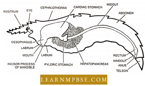
- Oesophagus. It is a short but broad tube which runs vertically upwards from the buccal cavity to the cardiac stomach. Internally the thick muscular walls of the oesophagus are thrown into four prominent longitudinal folds which project into its lumen.
- Due to these folds, the lumen of the oesophagus assumes a star-shaped appearance.
- Stomach. It is the largest part of the alimentary canal. It occupies most of the cephalorhoracic cavity. The stomach lies buried in the hepatopancreas, and it is covered over by the hepatopancreas laterally, posteriorly and ventrally.
- The stomach is divided into two unequal chambers: the cardiac stomach and the pyloric stomach.
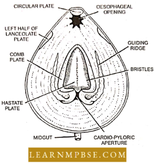
Cardiac stomach. It is a bag-like anterior chamber of the stomach. The internal chitinous lining (i.e., intima) of the cardiac stomach is thin and produced into numerous inconspicuous longitudinal folds covered by minute bristles. The wall of the stomach is supported by several calcified cuticular plates such as follows :
- Circular plate
- Lancevlate plate
- Hastate plate
- Combed plate
- Lateral grooves and groove plates
- Guiding ridges
Pyloric stomach. It is a very small narrow posterior chamber of the stomach. It lies below the posterior end of the cardiac stomach. Its lateral walls are thick and muscular.
- They divide the lumen of the pyloric stomach into a small dorsal chamber and a large ventral chamber. Both chambers are connected by a short narrow vertical slit-like apparture.
- Midgut It is a long, narrow, straight and slender tube which runs from the posterior part of the cephalothorax to the sixth abdominal segment. The midgut lies above the mass of the ventral abdominal muscles.
Hindgut. It extends from the posterior end of the midgut to the anus. It forms the shortest part of the alimentary canal. Anteriorly, the hindgut is swollen to a thick muscular sac, called the rectum while the posterior part is narrow and tubular which opens to the exterior through the. anus.
The rectum bears several thick longitudinal folds which project into the lumen of the hindgut. The rectum is lined internally by the chitinous cuticle.
Hepatopancreas. Die digestive gland, liver or hepatopancreas is a large bilobed, compact, orange-coloured gland which fills the cephalothoracic cavity. It surrounds the stomach and the anterior part of the midgut. Developmental!)’ it is formed from one pair of lateral tubular outgrowths, the hepatic caeca, of the midgut.
- The hepatopancreas of an adult consists of numerous glandular tubules which are branched in a resinous manner. These branches are held together by the connective tissue to form a compact mass. The wall of the tubules is formed of a single layer of columnar epithelium.
- This epithelium contains the following four types of cells. 1. granular cells; 2. ferment cells; 3, hepatic cells and -I. basal or replacing cells. All the epithelial cells rest on a basement membrane.
- Functions. The hepatopancreas functions like the liver, pancreas and small intestine of higher animals. It performs the following functions :
- Like the pancreas, it secretes digestive enzymes for the digestion of carbohydrates, fats and proteins. 2. Like the small intestine it absorbs digested food.
Like the liver. il serves as an important storage organ. It stores glycogen, fat and calcium. Some intracellular digestion also exists in its cells.
Food and Feeding: Polaemon is a nocturnal and omnivorous animal. It feeds on algae, mosses and other aquatic weeds. Occasionally, it feeds on small insects, snails, tadpoles, fishes, etc. and debris from the bottom.
The food is swallowed into the buccal cavity with the help of 1st pair of maxillipeds, maxillae and maxillulae. Inside the buccal cavity, food is masticated by the molar processes of mandibles.
Animal Nutrition And Digestive System NEET Notes
Digestion, Absorption and Egestion:
In Palaemon, the digestive process commences in the cardiac stomach.
- The hepatopancreas secretes enzymes into the pyloric stomach, from whence they travel to the cardiac stomach to combine with the food.
- The cardiac stomach alternately contracts and expands, facilitating the churning and enzymatic digestion of food.
- The undigested meal, containing unabsorbed material, ascends the dorsal chamber and proceeds into the midgut for absorption.
- The remaining digested food is absorbed in the midgut, while the residual faecal matter is sent to the hindgut and ultimately ejected through the anus.
Animal Nutrition NEET Notes
NEET Biology Animal Nutrition and Digestive System Of Vertebrates
The gut is divided only into functional parts not based on origin. The major parts are the mouth, pharynx, oesophagus, stomach, small intestine, large intestine and anus (or cloaca).
- It is longer in herbivores than carnivores.
- The major glands associated with it are the liver and pancreas.
Anatomy of Gut
Frog: Salivary glands are absent in frogs.
- The oesophagus is a short tube as a frog has no neck and thorax.
- The stomach is present in the middle of the body cavity and begins from the anterior part as a straight tube-like structure. Its posterior narrow pylorus makes ‘U with the duodenum, which lodges the pancreas.
- The small intestine is about 1-1.5 feet long.
- The large intestine is represented only by the rectum which opens into the cloaca.
Rabbit (Mammals) - The oesophagus is a long tube as that passes through the neck and thoracic cavity.
- The stomach is present in the left side of the abdominal cavity as a transverse bag-like structure and is roughly J-shaped.
- ‘U’ is formed by the duodenal loop only.
- The small intestine is about 4-5 feet long (giraffe = 250 ft).
- Sacculus rotundas act as a junction from which the caecum arises as a separate branch of the large intestine, as the site for cellulose digestion.
- Colon (50 cm) and rectum (75 cm) open into anus.
Ruminants. (for example cattle, sheep, goats, camel and deer) have a compound stomach with the following four chambers:
- Rumen First and the largest part for the storage of food and digestion of cellulose by symbiotic bacteria living here (Kumcnococcus).
- The reticulum (honeycomb) next chamber is also the site for cellulose digestion.
- Omasum (psalterium)-concentrates the food by absorbing H2O and H3CO, (absent in camel and deer).
- Abomasum or true stomach-last part is the secretion of gastric juice and digestion takes place here only. Camel’s rumen and reticulum have many diverticula (water pockets) but these do not store water.
Camel can live without water for two weeks. Its hump is a store of fats. For every 100 gms of fat metabolized, it produces 120 ml of H2O.
NEET Biology Digestive System Notes
NEET Biology Animal Nutrition and Digestive System Parts Of the Digestive System
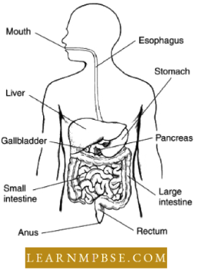
Parts of the alimentary canal of man.
- Buccal Cavity
- Pharynx
- Oesophagus
- Stomach
- Small Intestine (Duodenum Jejunum, and Ileum)
- Caecum and
- Large Intestine (colon and rectum). It terminates at the anal opening.
Digestive Glands
- Salivary glands
- Gastric glands
- Liver
- Pancreas
- Intestinal glands
Mouth Cavity: The mouth (oral or buccal cavity) is formed by cheeks, hard and soft palates and tongue.
- The vestibule of die oral cavity is bounded externally by cheeks and lips and internally by gum and teeth.
- The roof of the buccal cavity is the palate that separates it from the nasal chamber, consisting of hard and soft palate. The mucous epithelium has thick transverse folds called palatine rugae (thick in the case of carnivorous animals).
- The terminal part of the soft palate hangs a fleshy ‘V’ shape in the throat called the uvula.
On either side of the uvula, tonsils are present that are made of lymphatic tissue.
Tongue: It is a thick, musculo-sensory organ.
- Attached to the floor of the mouth by a soft ligamentous fold, the frenulum.
- Covered by the mucous membrane of thick stratified squamous epithelium with papillae of 4 types.
- The tongue is made up of striated muscles (voluntary).
- The tongue possesses Nuhn’s glands (glandular lingual autistics).
- Taste receptors (taste buds) are found mainly on the lingual papillae of the tongue.
- Filiform: Most abundant short filamentous (absent in Rabbit),
- Fungiform: Small mushroom-shaped on the upper part with taste buds.
- Foliate: Small leaf-like folds on the lateral side.
- Circumvallate: Largest cup-like, on the posterior part.
NEET Biology Digestive System Notes
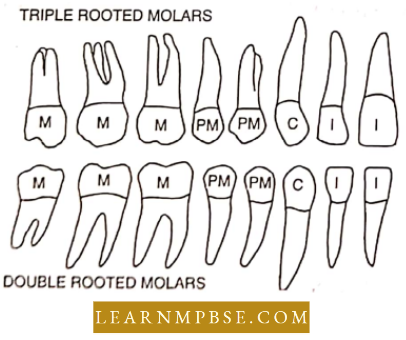
NEET Biology Animal Nutrition and Digestive System Dentition and Structure of Tooth
Originated from ectoderm and mesoderm.
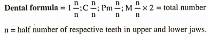
Dentition. The teeth are cone-shaped structures present in the mouth cavity. A full set of teeth in adult numbers 32 and child 20 represented by dental formula 2/2; c 1/1/; pm 2/ 2; m3/3 = 32 and 2/2: c 1/1: pm 0/0: m 2/2 = 20 respectively, as the teeth are heterodont initial represented by i-incisor. c-canine, pm-premolars and m-molar. The teeth of man are thecodont, and diphyodont. heterodont and bunodont.
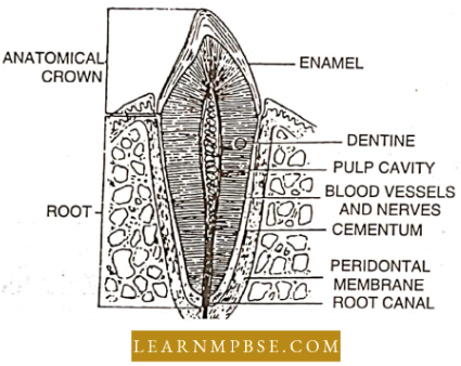
Structure of tooth. The tooth is divisible into three parts-root, neck and crown. The root is lodged in the socket or alveolus of the jaw bone and is firmly fixed by a cement-like substance called cementum. The bulk of the tooth is composed of a hard ivory substance called dentine.
It encloses a pulp cavity containing soft pulp composed of adipose tissue, blood vessels and nerve fibres. The pulp cavity is lined by a layer of odontoblasts. The neck part of the tooth is firmly held by fleshy gum. The crown is covered by the hardest substance called enamel. The teeth help in cutting and mastication of food.
NEET Biology Animal Nutrition And Digestive System Revision Notes
NEET Biology Animal Nutrition and Digestive System Types of Teeth
- Acrodont dentition. When the teeth are not embedded in sockets they are part of some bone as maxillary teeth and vomerine teeth of frogs.
- Thecodont dentition. When teeth are separate entities and are embedded in the teeth sockets as in mammals and crocodiles.
- Diphyodont dentition. When two sets of teeth are produced in the lifetime i.e. milk teeth and permanent teeth, as in Mammals.
- Polyphyodnnt dentition. When teeth can be replaced many times in life as a frog.
- Homodont Dentition (Isodont). When teeth are alike as in frog.
- Heterodont Dentition . When there are different types of teeth present, like incisors canines, premolars and molars as in Mammals.
- pleurodont Dentition. When the sides of teeth are fixed over the lateral surface of jaws as in reptiles.
- Bunodont Dentition. When there are low cusps present made by ridges of the teeth as in man,
- Solenodont. When the cusps are present as in sheep, etc,
- lophodont. When cusps fuse into transverse ridges as in elephants.
- Secodont. In carnivores such as cats. dog. lion. etc. cusps are pointed and are used in cutting.
- Crocodilian Teeth. Among the reptiles, crocodilians are exceptional in having theodont teeth. This suggests an evolutionary relationship between the reptiles and mammals.
- Carnnssial Teeth. In many carnivores (dogs, cats), the last upper premolar and the first lower molar are modified for shearing flesh. They are large and bite on each other like a pair of scissors. They are also called sectorial or shearing teeth.
- Eye Teeth. The upper canine is called eye teeth.
NEET Biology Animal Nutrition And Digestive System Important Points
NEET Biology Animal Nutrition and Digestive System Pharynx
The pharynx is about a 12 cm long vertical canal beyond the soft palate. The food and air passages cross here. The pharynx may be divided into 3 parts: nasopharynx, oropharynx and laryngopharynx.
Function. The function of the pharynx as a part of the digestive tract is merely to serve as a passageway for food from the oral cavity to the oesophagus. It has in its walls the voluntary muscles which start swallowing movements.
NEET Biology Animal Nutrition and Digestive System Oesophagus
Esophagus:
The esophagus is a 25 cm long, narrow, muscular, straight tube lined with stratified squamous epithelium containing mucous glands.
- It descends down the neck posterior to the trachea, traverses the thorax behind the heart, and passes through the diaphragm into the abdomen.
- Here, it sharply curves to enter the stomach. This bend is a mechanism to prevent the reflux of stomach contents into the oesophagus.
- Longitudinal folds maintain the cavity in a nearly closed state, except during the act of swallowing food.
- This monitors the air intake during respiration; the upper section of the oesophagus has striated muscle, the middle section contains a combination of striated and smooth muscle, while the lower section consists solely of smooth muscle.
Functionality:
The esophagus transmits food via peristalsis from the pharynx to the stomach.
Human Digestive System NEET Biology
NEET Biology Animal Nutrition and Digestive System Stomach
This stomach is wide, and J-shaped. distensible, muscular sac plated obliquely on the left side in the upper part of the abdomen just below the diaphragm. It is about 30. cm long and 15 cm wide. It has a greater curvature and a lesser curvature along its lower and upper sides.
The stomach is the most dilated part of the alimentary canal. In mammals, it is divided into three regions :
- Cardiac stomach- Anterior
- pyloric stomach-Posterior
- Fundic stomach-Middle
The narrow central end of the stomach is called the pylorus.
- The fundic part of the stomach is absent.
- An empty stomach is lined with folds called rugae.
- Ruminant animals such as cattle, buffalo, sheep, goats and camel have a compound stomach.
- compound stomach consists of four chambers, rumen, reticulum, omasum and abomasum.
- Some ruminants like camel and deer do not have omasum.
- Rumen is the largest and first of (ho four chambers.
- The rumen and reticulum are the sites of cellulose digestion these harbour numerous bacteria and protozoa w which carry out extensive fermentation of cellulose.
- Cyclostomes and lobed fishes do not have stomachs.
- Omasum concentrates the food by absorbing water and bicarbonates.
- The fourth chamber, abomasum is the (rue stomach as it secretes gastric juice and HCL.
- From the abomasum, the food passes to the small intestine.
- Temporary storage of food
- Mechanical breakdown of food
- Secretion of digestive juices.
- Partial digestion of food.
NEET Biology Human Digestive System Notes
NEET Biology Animal Nutrition and Digestive System Intestine
The intestine is quite longer in mammals, about 13 feet in rabbits, and 22 feet long in men because such a length increases the scope of food absorption.
- The intestine is usually longer in herbivores.
- The wall of the intestine is provided with only involuntary muscles.
- Small Intestine. The small intestine is a narrow tube, about 6 metres long in a living adult. It is the longest part of the alimentary canal. It comprises three parts: duodenum, jejunum and ileum.
- duodenum. It follows the stomach. It is somewhat C-shaped and about 25 cm. long.
- It receives the hepatopancreatic ampulla of the hepatopancreatic duct formed by the union of the bile duct and pancreatic duct.
- Jejunum. The jejunum is the middle part of the small intestine. It follows the duodenum and is about 2-4 metres long. Its wall is thicker and more vascular than that of the ileum.
- Ileum. The ileum forms the lower part of the small intestine. It is about 3-6 metres long and opens into the large intestine. Its wall is thinner and less vascular than that of the
Functions. The small intestine serves 2 main functions: completion of digestion and absorption of digested food. It also secretes some hormones such as cholecystokinin, secretin, duocrinin, and villikinin. entcrocrinin and enterogastrone.
NEET Biology Animal Nutrition and Digestive System Small Intestine
- In men, there is a common opening of the bile duct and pancreatic duct.
- Behind the duodenum is the jejunum, followed by the ileum.
- The lining of the small intestine bears a series of transverse folds called plicae circular or valves of Kerkering.
- Their internal lining is raised into innumerable minute finger-like processes called villi.
- In the wall of the small intestine, lymphatic tissues arc present collated Pcycr’s patches.
- Peyer’s patches are groups of lymph nodules that are most numerous in the ileum. They produce lymphocytes.
- The distal end of the ileum is expanded to form a small dilated spherical sac called sacculus rotundas in rabbits.
- The ileum opens into the caecum through an ileo-caccnl valve. large Intestine
- The large intestine is shorter than the small intestine. It is called the large intestine as it is wider than the small intestine. It is arranged around the mass of the small intestine.
- It is about 1-5 metres long. It lacks villi and microvilli. It shows three regions: caecum, colon and rectum.
- The caecum is very large and spacious in herbivores such as rabbit, horse and ass.
- The caecum of a rabbit is a thin-walled tube with peculiar external spiral constriction, which marks the presence of an internal spiral valve.
- Digestion of cellulose in rabbits takes place in caecum (Stomach in ruminants).
- Distally caecum terminates in a small, narrow, thick-walled tube vermiform appendix.
- But in man, the caecum is a reduced small pouch-like part below the opening of the ileum into the large intestine.
- In man, attached to the caecum is a twisted, coiled tube, measuring about 3 inches in length, called the vermiform appendix.
- The colon is thicker than the small intestine and thinner than the caecum. Constrictions of its wall form a series of small pockets called haustra.
- The haustra are arranged on either side of three median longitudinal muscle cords of the wall called taeniae.
- The colon of man is divided into ascending, transverse, descending and pelvic (sigmoid) portions.
- Mass peristalsis is initiated in the colon about half an hour after taking food.
- The pelvic colon continues into the rectum.
- The rectum is the last part of the alimentary canal, in man it is about 7-8 inches long.
- The terminal one inch of the rectum is called the anal canal and its exterior opening is called the anus.
- The Rectum of a rabbit is the narrow terminal part with faecal pellets present inside giving it a beaded appearance.
Human Digestive System NEET Biology
Functions. The large intestine mainly aids in the absorption of water; formation, temporary storage and elimination of faeces; and production of mucus for lubrication of mucosa.
- It also plays some role in digestion, absorption and excretion. The colon bacteria produce vitamins B and K which are absorbed.
NEET Biology Animal Nutrition And Digestive System Important Points
NEET Biology Animal Nutrition and Digestive System Digestive Glands
Salivary glands in humans 3 pairs in man, four pairs in rabbit, 5 pairs in rat and absent in frog. These are compound tubular-alveolar types.
Parotid gland
- It is present near the ear (pinna).
- It is the largest salivary gland but produces only 29% of saliva.
- It contains the enzyme- Ptyalin (a-amylase). Ptyalin is lacking in predators, Example: Lion and Tiger.
- The viral infection of these glands causes Mumps.
- The secretion of these glands is poured into the buccal cavity through Slenson’s duct.
Submaxillary (submandibular) gland
- It is present in the lower side of the upper jaw.
- It produces- 70% of saliva secretion.
- Its secretion reaches the buccal cavity through Wharton’s duct.
Sublingual gland
- It is present below the tongue.
- It is the smallest salivary gland and produces only 5% of the secretion
- Its secretion passes through the Duct of Rivins.
The salivary glands are absent in fishes, amphibians and aquatic mammals,
Saliva -1.5 lit/day
- pH – 6 to 7 (slightly acidic), on standing/heating it releases CO2 and becomes alkaline.
- The mucus of saliva helps in lubrication
- Saliva also causes the denaturation of raw proteins without hydrolyzing them.
- Thiocyanate ions, present in saliva, act as an antimicrobial agent to prevent infection by the microbes that often enter along with the food.
- Saliva also has lysozymes.
- Ptylain of saliva acts on boiled starch and converts it into maltose (a disaccharide).
NEET Biology Animal Nutrition And Digestive System Important Points
NEET Biology Animal Nutrition and Digestive System Liver
It is the largest sized, reddish-brown gland of the body. It is present in the posterior concavity of the diaphragm in the right upper part of the abdomen.
- The liver is a multilobulated gland. It is formed of two main and two small lobes. Two main lobes are the larger right and smaller left lobes, while two small lobes are the quadrate and caudate lobes.
- Each liver lobe is formed of hexagonal lobules surrounded by a connective tissue sheath called a Glisson capsule.
- Present on the lower surface of the right liver lobe, there is a thin-walled, pear-shaped sac, called the gall bladder. It stores the bile (about 60 ml) secreted by the liver.
- Bile is drained from the liver by a bile duct which is formed by the joining of a cystic duct from the gall bladder and a common hepatic duct from different liver lobes.
- Just near the duodenum, the bile and pancreatic ducts join to form the hepatopancreatic duct. The opening of the hepatopancreatic duct in the duodenum is guarded by a sphincter of Oddi. Daily secretion of bile is about 600-1200 ml.
- Bilirubin is formed by the daily destruction of haemoglobin due to the death of 1% RBC every day.
- The human liver weighs about 1.5 to 2 kg. and is divided into 3 lobes
- Glisson’s capsule, connective tissue sheath around each lobule, is a characteristic feature of mammals.
- Kupffer’s cells are large phagocytic cells to remove unwanted substances or foreign material from the liver hence, also known as hepatic macrophages.
- Hepatic capillaries arise from the cell level and unite to form the main hepatic duct emerging from each lobe to join the bile duct (ductus choledocus) which is also joined by the Cystic duct emerging from the gall bladder.
- The cystic duct conducts bile to the gall bladder.
- Opening of the bile duct in the duodenum is guarded by the sphincter of Oddi.
- The gall bladder is a separate part associated with the liver for the storage and concentration of bile.
- The gall bladder is a sac located along the underside of the liver.
- The gall bladder is absent in birds, rats, whales and horses.
- Gall bladder stores bile.
- Functions of Liver. The liver performs a variety of functions like synthesis, interconversions, storage, and secretion of various substances as follows:
- Glycogenesis. Extra glucose is converted to glycogen with the help of insulin and stored in this form either in the liver or in muscles.
- Glycogenolysis. Glycogen is converted into glucose when its level falls (below 80 mg/ 100 ml) in the blood. This is influenced by the glucagon hormone.
- Glucogenesis. Synthesis of glucose from other carbohydrates.
- Gluconeogenesis. Synthesis of glucose/carbohydrates from protein and lipids (non-carbohydrate sources)
- Lipogenesis. Extra protein and carbohydrates are converted into lipids for storage in adipose connective tissue.
- Deamination of protein. If protein is used for energy production, the NH2 group is removed from amino acid as NH2, the end product of keto acid, enters the Krebs cycle.
- Ornithine Cycle. The chemical part of excretion, NH3 is converted into urea in a cyclic chain of reactions in which ornithine plays a pivotal role.
- Cori Cycle. Lactic acid formed in muscle is convened buck In glycogen. Synthesis of substances like.
- VitA from carotene.
- Vit-D front cholesterol or ergocalciferol.
- Plasma protein (Albumin, globulin. clotting proteins). heparin.
- Angiotensinogen is an osmoregulatory substance in the blood.
- Haemoglobin, formation of UHC in frog and embryonic mammals.
NEET Biology Human Digestive System Notes
Somatomedin, a growth-promoting factor also called IGF (insulin-like growth I actor) Detoxification of the following substances take place in the liver
- Indole, skulls. cresol. phenol, alcohol, prussic acid and other toxic substances formed during digestion or obtained in blood.
- Excess alcoholism affects the liver by overburdening work and fat deposition.
- It converts alcohol to aldehyde which promotes lat deposition and causes puffiness and liver cirrhosis.
- Storage of the following substances takes place in the liver
- Glycogen, Vitamins like VitA, VitD, Vitk, VitB12 folic acid etc. Fe and Cu.
- Water, lymph and blood, (about 1/5th of total blood).
- By storing blood and water, the liver controls the volume and viscosity (concentration) of blood.
- By generating and consuming heat in a different chemical reaction it acts as a thermoregulatory organ.
Secretion of Bile
- Bile is a yellowish watery fluid, concentrated in the gall bladder, contains 85% water; 6% organic salt of bile acids (sodium glycocholate and sodium taurocholate); 1% inorganic salt (bicarbonate, carbonates and chlorides of Na and K); 1.5 to 2% lipid (cholesterol, lecithin); 3% mucin and bile pigment (bilirubin and biliverdin).
- Extra cholesterol and bilirubin are eliminated from the body through the gut.
- An increase in bilirubin level beyond normal (0.1 to 0.9 mg/100 ml.) in the blood is called jaundice. This happens if hepatic cells become sick due to viral or bacterial infection obstruction or lesion,
Gastric glands
- Gastric glands are microscopic and present in the walls of the stomach. These glands secrete gastric juice,
- A gastric, gland. has three types of cells.
- Mucous cells,- Secreting mucus
- Chief or peptic cell – Secreting pepsinogen zymogen cells and prorenin
Oxyntic. cells or parietal cells – Secreting HCL
Pancreas
The pancreas is the second largest gland in the human body. It is yellow-coloured, 12-15 cm long and a compound racemose gland. It is present in the loop of the duodenum. It is formed of head, body and tail.
- Pancreas is a heterocrine gland. Its exocrine part is formed of a large number of lobules or acini. Each acinus consists of several glandular cells which secrete the pancreatic juices (pH 8.8). Pancreatic juices are drained by a pancreatic duct which joins the bile duct as described in the liver. About 1500 ml of pancreatic juice is secreted per day.
- The rest part remains filled with areolar connective tissue known as stroma.
- Pancreatic secretion water-99%
- Inorganic – 0.5% (High amount of NaHCO3)
- Organic – 0.5% Mostly enzymes
Intestinal glands
- They are present in the wall of the intestine. These glands secrete intestinal juice (succus entericus)
- The wall of the duodenum contains intestinal glands (crypts of Lieberkuhn) and characteristic duodenal glands or Brunner’s glands produce alkaline mucus.
- In between the villi, the mucous membrane of the small intestine is folded forming intestinal glands or crypts of Lieberkuhn.
- The intestinal juice or succus entericus is secreted by the crypts of Lieberkuhn.
- Intestinal villi are more numerous and larger in the posterior part of the small intestine than in the anterior part because there is more digested food in the posterior part.
NEET Biology Human Digestive System Notes
NEET Biology Animal Nutrition and Digestive System Histology Of Wall Of Gut
- Histology of wall of gut from outer to inner side.
- Serosa – Peritoneum and Connective tissue Muscular Layer – Outer Longitudinal and inner Circular (Stomach – oblique muscles additional layers)
- Sulunueosa – Connective tissue Mucosa
- Muscularis mucosa
- Lamina propria (reticular connective tissue)
- Surface epithelium.
- Mouth and Pharynx
- Lined with the stratified squamous epithelium of ectodermal origin, hence representing the stomodaeal part. Possesses a striated muscle layer, hence the movement of food here is voluntary.
Oesophagus
- All four layers are typical as it serves only as the passage.
- Its thoracic part is without a serosa layer which shifts to form the mediastinal wall
Stomach
- Thick and highly distensible wall, when empty it forms inner longitudinal folds, rugae.
- The muscular layer is the thickest of all parts with the third and innermost oblique muscle layer.
- Submucosa has no speciality.
- Mucosa forms glands by invaginating into the submucosa and has three types of cells :
- Mucous (Goblet) cells secrete mucin
- Oxyntic (Parietal) cells secrete HCL
- Chief cells (Peptic or zymogenic) cells secrete enzymes.
- Cells of mucosa are secretory but not absorptive.
Enzymes In Digestion NEET
Small Intestine
The muscle layer is typical (no speciality).
- Submucosa is the most developed and specialised of all parts with the following structures :
- A highly organised system of blood capillaries up to villi for the transportation of absorbed food.
- Fine lymphatic channels (lacteals) are extended up to villi for absorption and transportation of lipids.
- The muscle layer (muscular mucosa) is present just behind the mucosa for the movement of villi.
- Two nerve plexuses of the autonomic nervous system (ANS) are present.
- The lymph nodes and Peyer’s patches are present which act as biological filters.
- Submucosa and mucosa together form inner transverse finger-like folds to increase the surface area of absorption. In mammals, the primary folds are called folds of Kerkering (or valvulae connivance or plica circular), which bear true villi as secondary folds.
- Folds of Kerkering increase surface area of absorption by 3 times, villi increase by 10 times and microvilli of mucosal cells increase by 20 times, thus total increase is by 600 times which comes to about 250 mtr2 in man.
- Mucosa is the main functional layer for digestion and absorption with brush border cells both secretory and absorptive. Mucosa forms glands embedded in submucosa are:
- Brunner’s gland (duodenal gland). Present mainly in the duodenum; secretes mucus; absent in the frog.
- Crypts of Lieberkuhn. Small flask-shaped gland in the crypts between two villi.
- Cells of Paneth in it secrete digestive enzymes, called succus entericus
Large Intestine: Thin-walled and transparent, divided as caecum, colon, and rectum.
Colon
- No secretion, no glands at all.
- There are three inner longitudinal folds called taeniae – specialized structures for the absorption of water.
- The transverse membranous foils between two taeniae form a series of pockets, dentistry, highly developed in desert or dryland animals like rabbit, pont. camel etc. but, less developed in man
Enzymes In Digestion NEET
NEET Biology Animal Nutrition and Digestive System Rectum
- Has six or more folds called rectal papillae for the absorption of water,
- Goblet cells secrete mucus to facilitate defecation.
- Physiology of Digestion
- Digestion is the process of conversion of complex food into simple (absorbable) form, lists involve two parts :
- Mechanical part. Cutting and chewing of food to increase its surface area for enzymatic action,
- Chemical part. Enzymatic hydrolysis of food i.e. breaking tit molecular level i.e. actual digestion.
- It could be both extracellular and intracellular, but secures, optimal efficiency of extracellular digestion.
- Discovered by Beaumont (1833).
- It begins from the mouth and continues up to the small intestine, in herbivores digestion also takes place in the caecum but with the help of bacterial enzymes.
NEET Biology Animal Nutrition and Digestive System Absorption Of Food
Talcs place, mainly jejunum and ileum through villi and microvilli by both active and passive preveses. It first enters mucosal cells and then passes into the submucosa.
- No absorption occurs in the stomach except that of ethanol (alcohol) and aspirin.
- Water is absorbed in the small intestine and large intestine.
Absorbed food is transported through two pathways: the hepatic portal system, directly to the liver and through the lymphatic channel it is drained into subclavian veins via the thoracic duct.
Digestive Enzymes And Their Functions NEET Study Material
Absorption of Glucose and Amino acids
- Both are absorbed mainly by active transport as this process is many thousand times faster than passive transport.
- Entry of glucose in the cell is coupled with Na+ where it immediately gets phosphorylated.
- Galactose is absorbed like glucose, and some disaccharides are absorbed directly.
- Fructose and mannose are absorbed by facilitated diffusion.
- From submucosa, these pass out through blood capillaries.
Absorption of Lipid: Glycerol and fatty acids in the cell generally combine to form lipids again. Thus lipid is absorbed in both forms :
Directly as lipid
- Lipid enters mucosal cells as chylomicrons and micelles by pinocytosis in the crypts between villi.
- Chylomicrons are lipid droplets of < I pm diameter formed by emulsification. With non-lipid substances like salts, amino acids etc. it forms micelles.
- In submucosa, it enters lacteals and moves with lymph.
- As glycerol and fatty acid
- Glycerol is soluble in both the aqueous and lipid phases and hence easily passes through the cell membrane.
- Small soluble fatty acids enter the cell as the membrane component, while large insoluble fatty acid after combining with (bile) forms a soluble micelle.
Animal Nutrition And Digestive System NEET Chapter Summary
NEET Biology Animal Nutrition and Digestive System Egestion (Efaecation or Defecation)
- It is a tile process of elimination of faeces.
- Slercobilin causes the brown/yellowish colour of faeces.
- The foul odour of faeces is due to indole and skatole funned by decarboxylation of amino acids by bacteria in the colon.
NEET Biology Animal Nutrition and Digestive System Components Of Foods
The food of animals is chiefly formed of six kinds of components
- Carbohydrates
- Proteins
- lipids
- vitamins
- Water
- Minerals.
Out of these food components, carbohydrates, proteins, and fats are called macronutrients or proximate principles of food, while vitamins and minerals are called micronutrients or protective principles of food.
Proteins
- Proteins are major building blocks as 75% of our body consists of proteins. All enzymes, O. earning pigments, antibodies, most hormones, arc proteins.
- The chief source of proteins is milk. egg. fish. meat, pulse and cereals. Its requirement is more for growing children and pregnant and lactating mothers.
- Proteins are more important for anabolism than catabolism Calorific value of proteins is 5.65 K cal and 20 amino acids, linked together by peptide bonds, make all different proteins.
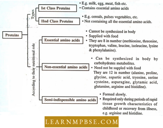
Eight are considered essential (which cannot be synthesized in the body and lias to be taken in diet), and 12 are non-essential (which can be synthesized in the body) which 2 are considered semi-indispensable because they may be synthesized in tissues but not at adequate rates to support growth in younger human individuals.
Digestive Enzymes And Their Functions NEET Study Material
NEET Biology Animal Nutrition and Digestive System Carbohydrates
- Chemical composition. Carbohydrates are polyhydroxyaldehydic or ketonic organic compounds. These are usually formed of C, H and O in the ratio of 1:2:1 with only a few exceptions.
- These are called carbohydrates because there is generally one water molecule for every carbon atom. The general formula is :
- Cn(H2:0)n where n is several carbon atoms, so arc called “hydrates of carbon” The Name of carbohydrates ends with the suffix – ose.
- Sources. Main sources of carbohydrates are potatoes (20%), fruits (banana – 20%, mango), cereals (rice – 23%. wheat – 57.3%), sugar, honey, sugarcane, sugarbeet, jam,’ bread, milk 4%) etc.
Daily requirements. On average, the need for carbohydrates for an adult person is 100 g per day. About 55-75% of total good calories should be in the form of carbohydrates.
- Of these. 80-85% should consist of easily digestible starch and dextrins. Requirements are more for mountaineers, athletes, labourers etc.
- Carbohydrates form about 1% of our body weight.
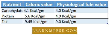
Functions. As Respiratory fuels. Carbohydrates, especially glucose, are the main respiratory fuels. About 60% of our total energy needs are provided by the breakdown of carbohydrates is 4.1 kcal of energy while the physiological fuel value of one gram of carbohydrates is 4.0 kcal (17 kJ) of energy.
- To provide energy, the glucose undergoes biological oxidation in the mitochondria (powerhouse) of the cell to produce about 36 or 38 molecules of ATP.
- So die theoretical recovery- of energy from one glucose molecule of 40%. The main reasons for the glucose being chief respiratory fuel are its presence in abundance and its easy oxidisability.
- C6H12O6+6O2 > 6CO2 + 6H2O + Energy
- So carbohydrates form the cheapest sources of energy.
- Monosaccharides as structural components
Ribose (Pentose sugar) is a component of RNA; conenzymes like NAD, FAD etc. and energy carriers like ATP, GTP etc.
- 2′-Deoxyribose (Pentose) is a component of DNA.
- Galactose is a structural component of the medullary sheath.
- As building blocks. Monosugars act as monomers for the formation of disaccha¬rides and polysaccharides.
- Reserve foods. There are two main polysaccharides which act as reserve foods e.g. starch is a storage polysaccharide of plants and is stored as granules in amyloplasts.
- Glycogen is the reserve food stored in the liver and muscles in animals.
- The energy requirement of the body Organisms requires energy continuously. When work is done, energy is being spent. This energy is required to keep all systems in the body functioning.
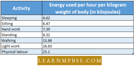
- A normal healthy man requires about 3000-kilo calories per day. Perhaps a sedentary worker needs a little less, say 2500 kilo calories hardworking labourer may need about 3500-kilo calories per day.
- (Note: The term Calorie (capital C) as used by nutritionists is equivalent to 1 kilo calorie (kcal)) The energy requirement is calculated as so many calories for an hour for a particular type of work.
- Basil metabolism. The amount of energy required by the body to maintain vital functions when no muscular work is being done and no food is being digested.
- Rasul metabolic rate (BMR). The amount of energy required daily by a person to maintain their basal metabolism is about 1,600 keal/day.
- Active metabolic rate (AMR). The amount of energy required daily by a person to maintain their high metabolic rate during heavy physical work is about 6,000 kcal/ day.
- Roughage. The indigestible fibrous material of food is called roughage For Example. cellulose of cell walls of plant material promotes peristalsis and checks constipation.
- Balanced diet. A diet which can provide materials for all the metabolic requirements of the body. Carbohydrates, proteins and fats should be nearly in the proportion of 4: 1: 1.
Lipids. Lipids comprise heterogenous organic compounds which are insoluble in water but are readily soluble in non-polar organic solvents like ether, chloroform, benzene etc. On hydrolysis.
- lipids yield fatty acids which are utilized by living organisms. In addition to fats, lipids include waxes, phospholipids, glycolipids and fat-soluble vitamins A, D, E and K).
Daily Requirement. About 50 gms. of fat are needed by man daily. Fats are digested slowly and delay the hunger sensation between meals. Our diet should contain less saturated fats, such as butter, ghee and hydrogenated vegetable oils, than unsaturated fats, such as simple vegetable oils.
- Excess of saturated fats increases the cholesterol and causes arteriosclerosis.
Sources. The sources of fats in our diet are vegetable cooking oil, vanaspati ghee, desi ghee, butter, cream, oil seeds and nuts, milk, cheese, mutton and eggs.
Enzymes In Digestion NEET
Uses. The fats also serve a variety of functions:
- fuel,
- reserve food
- insulator
- formation of cell organelles
- shock absorption.
Animal Nutrition And Digestive System NEET Chapter Summary
NEET Biology Animal Nutrition And Digestive System Vitamins
N.I. Lunin was the first to identify vitamins, and Funk was the first to use the term “vitamins.” The “vitamin theory” was introduced by Hopkin and Funk in 1912, which posits that deficiencies in vitamins are the cause of deficiency diseases.
- As growth and metabolic regulatory substances, vitamins are intricate organic compounds that are required daily in minute quantities.
- Green plants are the sole source of vitamin synthesis; consequently, animals are reliant on plants to meet their vitamin needs.
- The human body can store A, D, and ll,2 and produces vitamin D through the use of ultraviolet radiation of sunlight. Vitamins are named after the alphabets they represent, their chemical composition, and the deficiency diseases they prevent.
- Vitamins are compromised by excessive heating, frying, and low temperatures. It is feasible to synthesize vitamins, as their chemical composition is recognized. Vitamins are classified as either fat-soluble (A, D, E, and K) or water-soluble (B and C).
Animal Nutrition And Digestive System NEET Chapter Summary
NEET Biology Animal Nutrition and Digestive System Memory Points
- PFA Polyunsaturated fatty acids are found in abundance in corn oil and sunflower oil
- NiN National Institute of Nutrition
- Vit C was the first vitamin to be produced during the fermentation process using wild bacteria.
- Vit B12 was first to be isolated in 1948 from liver extract and during the production of antibiotics in fermentation.
- Within the body, vitamin B12 is produced by the microflora of the digestive system.
Excessive intake of vitamin A causes bone reabsorption and hypercalcaemia.
- An alcoholic is always deficient in Vit. C
- Vitamin B17 is a recently discovered vitamin with anti-cancer properties.
- Vitamin theory was proposed by Hopkins and Funk.
- Earlier known vitamin -Vitamin C
- Vitamin B6 is used in the treatment of tuberculosis
- Vit P Hesperdin is insoluble in water and is present in citrus fruits and green vegetables. It maintains blood capillary resistance. Its deficiency causes subcutaneous bleeding. ,
- Vitamins which are synthesized by the intestinal flora are vitamin K, Thiamine, Riboflavin, Pantothenic Acid, Niacin, Pyridoxine, Biotin and Folic acid.
- Vitamin B12 and alcohol. Alcohol interferes with the metabolism of B1 in the liver. Alcohol intake along with a deficiency of vitamin B1 can also cause brain cell damage. Therefore, regular alcohol drinkers should take vitamin B complex every day.
- Cod Liver oil is considered to be a rich source of vitamins A and D.
- Presently Vit B12 is produced directly during the courses of fermentation by propioni bacteria and certain strains of Pseudomonas.
- Vit B12 is found essentially in cereals, vegetables and brewer’s yeast. It was produced in 1938 by using microbes.
- Most of the B complex vitamins are coenzymes.
- Wheat grain contains – Vitamins such as thiamine, riboflavin, niacin, pantothenic acid, folic acid, and tocopherol (The richest source)
- Proteins & Minerals P, K, Mg, Mn, Ca, Na, Sn, Zn. & Fat.
- Destruction of vitamins. Overcooking, excessive alcohol, tobacco and coffee; certain medicines destroy vitamins
- Dietary deficiency of proteins leads to a fall in plasma proteins which are important for the retention of water in blood plasma by their osmotic effect.
- A fall in plasma proteins leads to filtering out excessive volumes of water from the blood to the tissues. Excessive accumulation of fluids in the body tissues causes oedema.
- Zinc is necessary to maintain normal plasma concentration of vitamin A.
Animal Nutrition And Digestive System NEET Chapter Summary
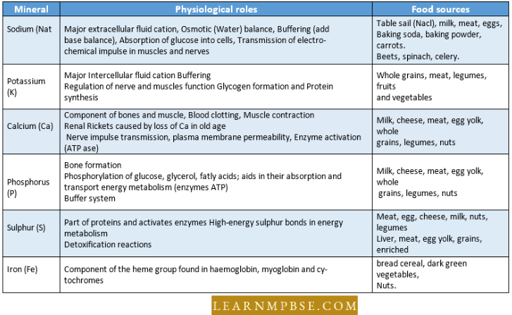
NEET Biology Animal Nutrition And Digestive System Physiological Role And Source Of The Important Minerals
NEET Biology Animal Nutrition and Digestive System Disorders Of the Digestive System
- Nausea- discomfort preceding vomiting.
- Anorexia – loss of appetite.
- In India, Orissa tops the list of states of people suffering from malnutrition.
- Enlargement of rectal vein which causes piles.
Dyspepsia. Indigestion due to defective diet.
- Pavlov pouch was fabricated by Pavlov to study the effect of feeding on gastric secretion.
- Peristaltic movements are low in the rectum.
- A hiatal hernia or diaphragm is the opening in the diaphragm. The part of the stomach is pushed into the thoracic cavity.
- Gastritis is inflammation of the gastric mucosa.
- A peptic ulcer is an erosion of the stomach or duodenal mucosa.
Nutrient Absorption In Small Intestine NEET Exam Preparation
Cirrhosis of the liver – The liver appears orange. It is due to the accumulation of bilirubin in the liver. Other substances are mixed with the yellow pigment (bilirubin), hence the liver appears orange.
- Hepatitis is inflammation of the liver.
- Pancreatitis is inflammation of the pancreas.
- Constipation is the infrequent passage of dry, hardened faeces.
- In ulcerative colitis the mucosal lining of the colon becomes ulcerated.
- Some people cannot digest milk and milk consumption in them causes diarrhoea and gas generation because they do not produce lactase. The lactose of the milk is fermented by intestinal microflora-producing gas.
- Removal of the stomach produces dumping syndrome.
- Treatment with all axons destroys 3 cells of pancreatic islets
- Treatment with CaC12 destroys cells of islets of Langerhans
- Abnormal metabolism of fats causes Gaucher’s disease (also called cerebroside lipidosis)
- PEM Protein Energy Malnutrition
- Kwashiorkor Marasmus
- FFA Free fatty acid,
- Avg. Indians Iwvo to obtain about 50% of their requirements of energy from early dratos, 33% from fats and 15% from protein.
- The vermiform appendix (1.. vermiformis, worm-like; appendix, attachment) is a short, thin outpouching of the caecum. It does not function in digestion, but like the tonsils, it contains numerous lymphatic nodules and is subjected to inflammation a condition called appendicitis.
- It is commonly detected in its later stages be pain. Rupture of the appendix can cause inflammation of the surrounding body cavity peritonitis.
- The substances which provide materials for growth, energy and maintenance are called nutrients.
- The process of procurement of nutrients is called nutrition.
- The mode of nutrition may be autotrophic or heterotrophic.
- Nutrients may be organic or inorganic.
- Proximate principles of food
- They are macronutrients
- These include carbohydrates, lipids and proteins.
- They are energy sources and help in carrying out organic functions.
- Protective principles of food
- They are micronutrients.
- These include water and minerals.
- They do not provide energy but help in carrying out various vital processes.
- About 20 vitamins are required in small amounts.
- The mode of nutrition in animals is heterotrophic which may be holozoic or saprozoic.
- Based on food habits holozoic animals are classified into herbivores, carnivores, omnivores, insectivores, sanguivores, detritivores, insectivores or fluid feeders.
- Ingestion is the intake of food.
- Microphagous animals. These animals feed on those solid particles which are too small to be captured singly. These animals possess different types of filtering devices. Therefore, the method of intake is also known as filter feeding.
- The water passes through the filters and food is retained (e,g., pseudopodia, cilia, flagella and setae in some crustaceans etc.)
- Filter feeders. They are aquatic animals and feed on tiny particles suspended in water.
- Examples. Lepas (Crustacean), Molluscs, Amphioxus. Herdmania.
- Macrophagous animals. These animals feed on those solid particles which are large enough to be captured singly. Filters are absent in these animals. The method of capturing food is different in different animals.
- Pseudopodia capture and ingest the food in amoeba. This process is called phagocytosis. Earthworms spreads its mouth cavity to capture the food. Coelenterates capture the food with the help of tentacles.
- Fish, amphibians, creeping animals and birds capture their food with the help of jaws, tongues and beaks and ingest it without chewing them. The food captured by most of the mammals is masticated before being swallowed.
- Their teeth as well as jaw bones and muscles are specially developed for mastica¬tion. In herbivores, the premolars and molars have predominant ridges on the crowns for effective grinding. The carnivores, however, have large and sharp canines for tearing the flesh of prey.
Mutualism. Two organisms living in association with each other derive nutrition from each other, in such case nutrition is called mutualism.
Examples of mutualism
- E.coli living in the intestine of man synthesises vita-min B12, which is utilised by man and E.coli receives, in return simpler food from the intestine of man.
- Rhizobium bacteria live in the nodules of leguminous plants.
- The cloacal thymus is the purely endocrine gland on the roof of the cloaca of the birds discovered by Fabricius.
- Mass peristalsis is initiated in the colon about half an hour after taking food.
- Riboflavin was called ‘yellow enzyme’ by Warburg and Christian.
Animal Nutrition And Digestive System NEET Chapter Summary
NEET Biology Animal Nutrition And Digestive System Quanta To Memory
Oesophagus. In Birds, the oesophagus is very long and often dilated in front of the sternum into a large, thin-walled crop to store food before digestion. Crop glands in both sexes secrete a nutritive fluid the pigeon’s milk, which is regurgitated and fed to the hatchlings.
- In the upper one-third of the oesophagus, only skeletal muscles are found.
- After the voluntary phase, swallowing of ‘bolus’ becomes an involuntary reflex triggered by tactile stimulation of the palate and pharyngeal wall controlled by the swallowing centre in the medulla and lower parts of the brain stem.
- In humans, more than one litre of saliva is secreted into the oral cavity.
- spite of the fact the mucus protects the stomach lining from being digested, it is completely replaced every 3 days.
- Each day, the stomach wall secretes about 3 litres of gastric juice.
- About every 20 seconds, the stomach contents are mixed by the churning action of smooth muscles. When an empty stomach churns, hunger pangs are felt.
- It takes about 2-6 hours after a meal for the stomach to empty.
- Amylases found in animals are called a-amylose, and those in plants are called /Lamylose.
- Stomach. Cyclostomes and the carp, Labeo, lack stomach.
- Water Cells. These are sac-like diverticula arising from the rumen and reticulum of the camel stomach. They contain food undergoing digestion and do not store water as commonly supposed.
- Camel can go without taking water for a long time as it can use metabolic water formed by chemical breakdown of stored glycogen and fat.
- Intrinsic Castle’s Factor. It is secreted by parietal cells of the gastric glands and aids in vitamin B]2 absorption. Lack of this vitamin makes the bone marrow unable to form sufficient red corpuscles, causing pernicious anaemia.
- Small Intestine. Final digestion of double sugars right on the surface of microvilli and of dipeptides on the microvilli and within the microvilli-bearing cells. Products of digestion, viz., glucose and amino acids, are released into the intestinal lumen.
- Paneth Cells. These occur at the base of the crypts of Lieberkuhn and secrete antibacterial lysozyme.
- Argentaffin Cells. These also occur at the base of the crypts of Lieberkuhn. They secrete some gastrointestinal hormones and neurotransmitters.
- Peyer’s Patches. These are white patches on the intestinal mucosa. They consist of lymphoid tissue packets with white corpuscles. They fight infection and are often called abdominal tonsils.
Enzymes In Digestion NEET
Large intestine. The vermiform appendix contains lymphoid tissue that neutralizes bacterial toxins.
- Some of the bacteria including Escherichia coli present in the large intestine or colon produce vitamin K, which is absorbed by the host and this is probably the main source of this vitamin for humans.
- Millions of cells lining the human stomach and intestine are abraded away and destroyed every time the food is digested, but cell division constantly regenerates the lining of the digestive tract.
- Every five days, the lining of the human small intestine is entirely replaced as a result of ongoing cell division.
- Parietal cells of the stomach give MCI and chief cells give pepsinogen.
- The surface of the upper gastrointestinal tract, oesophagus, and mouth have a much thinner, mucous cell layer than the stomach, which is why vomiting can cause, a burning sensation in the oesophagus or mouth.
- The vermiform appendix (L. vermiform’s, worn-like; appendix, attachment) is a short, thin outpouching of the caecum. It does not function in digestion, but like the tonsils, it contains numerous lymphatic nodules and is subject to inflammation a condition called appendicitis.
- It is commonly detected in its, later stages by pain. Rupture of the appendix can cause inflammation of the surrounding body cavity peritonitis.
- About 40-50 lakh villi are present in the human intestine.
- On the surface of each epithelial cell of villi about 650 microvilli are present, ‘
- The functional and structural unit of the liver is the liver lobule Part of the alimentary canal where NH3 (Ammonia) is produced during digestion is the caecum
- Bursa Fabrici. It is a blind sac on the cloaca in birds. It has lymphoid tissue involved in antibody production and in fighting invading bacteria. It is, therefore called cloacal thymus.
- Like the thymus, it is prominent in early life and tends to atrophy in the adult.
- The centre in the brain which controls hunger is known as the satiety centre. It is located in the hypothalamus of the brain.
- The chief function of the ileum is absorption.
- The most important functions of saliva are mechanical.
- Piscivorous animals feed on fish. ,
- Saliva and tears contain an antibacterial enzyme called lysozymes.
- Poison glands of snakes are modified labial glands homologous to parotid salivary glands.
- The liver of a rabbit is encapsulated by two sheaths – an outer membranous serous capsule consisting of a visceral peritoneum and an inner Glisson’s capsule of a thin layer of dense connective tissue. Glisson’s capsule is found in the liver of some mammals.
- The rumen, Reticulum, Omasum and Abomasum are the correct sequence through which food passes through a ruminant.
- Diastema is a toothless space in rabbits between the incisors and premolars.
- Fibres running between the periodontal membrane to the root of the tooth across the cement are known as Sharpey’s fibres.
- C-shaped duodenum is a characteristic of man. Reverse peristalsis or anti-peristalsis in the stomach and duodenum causes vomiting.
- The vomiting centre is the medulla oblongata of the brain.
- Some people suffer from “travelling sickness” on their way to a hill station in a bus, the sickness comprises nausea and vomiting, which are due to rapidly changing directions of motion of the body stimulating the receptors of the membranous labyrinth, from where the stimuli reach the vomiting centres in the medulla oblongata through the cerebellum.
- Peristaltic movements are the least in the rectum of the alimentary canal.
- In herbivorous animals, the small intestine is much longer (tadpole) than that of carnivorous animals (For Example. frog in proportion to body size)
- In rabbits, there are 4 pairs of salivary glands: Parotid, sub-axillary, sub-lingual and infraorbital (below the orbits and absent in man).
- Liver. It is relatively much larger in the foetus. It has a double blood supply: about 70% coming from the hepatic portal system and 30% from the arterial system.
- The liver of a rabbit is formed of five lobes: Right central (largest), left central, left lateral, caudate and Spigelian (smallest and absent in the liver of man).
- Faecal matter is solid in the descending colon.
- The lining cells of the small intestine have microvilli for absorbing digested food. The microvilli are not present in the embryo but develop after birth as the young one starts suckling.
- Once glycogen reserve is exhausted the adipose tissue (fat cells) comes to the rescue of the fasting person.
- Bp is absent in plants, but present in almost all animal tissues of which rich sources are liver, kidney, milk, etc.
- Tapeworms are said to be “Wallowers” because they absorb nourishment through their body surface.
- Rennin is secreted by young mammals. It is not secreted by adult mammals.
- The first pancreatic transplant took place in l%6 in the U.S.
- Bad breath is usually due to cavities in the teeth, and infection of the throat and nose.
- Many people are overfed but undernourished.
- Tea/coffee inhibits the absorption of iron from the diet. Prolonged consumption of tea or coffee after meals can lead to iron deficiency anaemia.
- The cells of the epithelial lining in a vertebrate stomach are not damaged by HC1 because of mucus secretion covering the epithelium.
- Under extreme demand of glucose (starving) even reserve proteins and fats are converted into glucose through a process of gluconeogenesis.
- Wound healing is enhanced by Vit-C.
- Lips (Labia Ora). Chelonians and birds lack lips. The upper lip of rabbits, hares, rats and squirrels has a median vertical cleft, the harelip, exposing the upper teeth. Some humans also have harelip as a congenital abnormality.
- It can be set right by a surgical operation. The upper lip and snout are prolonged into a long, thick, prehensile proboscis or trunk in elephants. Platypus and whales lack movable lips.
- Cheek Pouches. In some rodents (squirrel, rat) and certain old-world monkeys, the vestibule (space between cheeks and jaws) extends to form cheek pouches for temporary storage of masticated food.
NEET Biology Human Digestive System Notes
NEET Biology Questions For Competitive Examinations
Question 1. Protein/enzyme is absent in :
- Saliva
- Bile
- Pancreatic juice
- Intestinal juice.
Answer: 2. Bile
Question 2. Match the degestive juices listed under Column-1 with the enzymes given under Column-2; Choose the choice which gives the correct combination of the alphabets of the two columns.
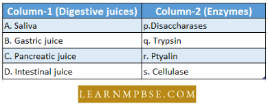
- A — r, B — s, C — p, D — t
- A — r, B — t, C — p, D — q
- A — s, B — r, C — p, D — t
- A — r, B — S, C — q, D — p.
Answer: 4. A — r, B — S, C — q, D — p.
Question 3. What should be taken to offset the deficiency of rhodopsin?
- Papaya and Mango
- Orange and Amla
- Watermelon and Strawberry
- All the above.
Answer: 1. Papaya and Mango
Question 4. Vitamins required for the development of erythrocytes are:
- D
- B12
- E
- K.
Answer: 2. B12
NEET Biology Human Digestive System Notes
Question 5. In the case of taking food rich in lime juice, the action of ptyalin on starch is :
- Enhanced
- Reduced
- Unaffected
- Stopped.
Answer: 2. Reduced
Question 6. Digestive juice contains catalytic agents called :
- Vitamins
- Hormones
- Enzymes
- Nitrates.
Answer: 3. Enzymes
Question 7. Bile acids are :
- Steroids
- Carbohydrates
- Modified proteins
- Vitamins.
Answer: 1. Steroids
Question 8.Which one correctly matched :
- Vit. E-Tocoferol
- Vit. D-Riboflavin
- Vit. B-Calciferol
- Vit. A-Thiamine
Answer: 1. Vit. E-Tocoferol
Question 9. Where does the conversion of harmful prussic acid into potassium sulphocyanide take place?
- spleen
- liver
- bone marrow
- lymph glands.
Answer: 2. liver
Question 10. Water is absorbed mainly by :
- Large intestine
- small intestine
- stomach
- pancreas.
Answer: 1. Large intestine
Question 11. The number of different teeth (incisors, canines, pre¬molars and molars) in each jaw are :
- 4, 4, 6. 4
- 4,2,4,6
- 4, 2, 5, 6
- 6, 2, 4, 4.
Answer: 2. 4,2,4,6
Question 12. “Your food shall be your medicine”. This quotation was given by :
- Newton
- Hippocrates
- Einstein
- Lenz.
Answer: 2. Hippocrates
Question 13. Term proteins were first used by :
- Berzelius
- Funk
- Kuhne
- Marconi.
Answer: 1. Marconi
Question 14. Gall baldder is absent in :
- cow
- pig
- horse
- cat.
Answer: 3. horse
Disorders Of Digestive System NEET Biology
Question 15. Vitamin K is necessary for:
- R.B.C.
- W.B.C.
- plasma
- production of prothrombin.
Answer: 4. production of prothrombin
Question 16. A person suffering from profuse bleeding after an injury. Such a person is deficient in which vitamin?
- Vit A
- Vit K
- Vit D
- Vit E.
Answer: 2. Vit K
Question 17. Which of the following is the largest part of a cow’s alimentary canal?
- Reticulum
- Rumen
- Omasum
- Abomasum.
Answer: 2. Rumen
Question 18. Absorption of glucose and amino acids occurs by:
- Passive absorption
- Active absorption
- Simple diffusion
- Facilitated diffusion.
Answer: 2. active absorption
Question 19. Magnesium is most abundant in :
- milk
- meat
- soybean
- fish.
Answer: 2. meat
Question 20. Deficiency of which of these causes anaemia?
- Fe
- Zn
- A1
- Mg.
Answer: 1. Fe
Question 21. Which one pair is not correctly matched?
- Vit Bp-Pernicious anaemia
- Vit B6-Beri-Beri
- Vit C-Scurvy
- Vit B2 -Pellagra.
Answer: 4. Vit B2 -Pellagra
Question 22. Lysozymes are found in :
- Mitochondria
- Tears
- Saliva and tears both
- Saliva.
Answer: 3. Saliva and tears both
Question 23. Just as hydrochloric acid is to pepsinogen so is :
- Enterokinase to trypsinogen
- Haemoglobin to oxygen
- Bile juice to fat
- Glucagon to glycogen.
Answer: 1. Enterokinase to trypsinogen
Disorders Of Digestive System NEET Biology
Question 24. Plasma protein also performs :
- Nutritive function
- Physioehemienl function
- Transportive function
- All the above three.
Answer: 4. All the above three
Question 25. Which is non-reducing sugar?
- glucose
- galactose
- mannose
- sucrose.
Answer: 4. sucrose
Question 26. Which is false for nutrition in Amoeba?
- pseudopodia feeder
- holozoic nutrition
- photonivorous
- Photoautotroph.
Answer: 4. photoautotroph
Question 27. Match the types of cells listed under column 1 with the secretions under column II. Choose the answer which gives the correct combination of the alphabet of two columns :
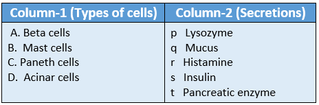
- A = s b = r ; C = p D = t
- A = q b = r; C = p D = t
- A – s b = q; C = p D = t
- A = t b = q; C = r, D = S.
Answer: 1. A = s b = r ; C = p D = t
Question 28. The lacteals are found in :
- Spleen
- Mammary gland
- Salivary gland
- Villi.
Answer: 4. Villi
Question 29. Number of teeth which grow twice are :
- 8
- 14
- 12
- 20.
Answer: 4. 20
Animal Nutrition And Digestive System NEET Notes
Question 30. Which of the following is not an insectivorous plant?
- Drosera
- Nepenthes
- Monotropa
- Utricularia.
Answer: 3. Monotropa
Question 31. Seaweeds are important sources of :
- Chlorine
- Fluorine
- Iodine
- Bromine.
Answer: 3. Iodine
Question 32. During digestion, lymphatics of the intestine become filled with fat globules giving a white colour to the lymph. This lymph is called :
- Cistron
- Chyle
- Chyme
- Bilirubin.
Answer: 2. Chyle
Question 33. Cattle fed with spoilt hay of sweet clover which contains dicumarol :
- Are healthier due to a good diet
- Catch infections easily
- May suffer from vitamin K deficiency and prolonged bleeding
- May suffer from Beri Beri due to a deficiency of B vitamins.
Answer: 3. May suffer from vitamin K deficiency and prolonged bleeding
Question 34. Duodenum has characteristic Brunner’s glands which secrete two hormones called :
- Prolactin, Parathormone
- Estradiol, Progesterone
- Kinase, Estrogens
- Secretin, cholecystokinin.
Answer: 4. Secretin, cholecystokinin
Question 35. The richest sources of vitamin B,2 are :
- Chocolate and green grain
- Rice and Men’s egg
- Carrol and chicken’s breast
- Goat’s liver and Spirulliiui
Answer: 4. Goat’s liver and Spirulliiui
Question 36. Which one of the following pairs is not correctly matched?
- Vitamin B1 = Beri-Beri
- Vitamin B2 = Pellagra
- Vitamin B12 = Pernicious anaemia
- Vitamin B6 = Loss of appetite.
Answer: 2. Vitamin B2 = Pellagra
Question 37. Which one of the following is the correct matching of a vitamin, its nature and its deficiency disease?
- Vitamin A – Fat-soluble – Beri-Beri
- Vitamin K – water-soluble – Pellagra
- Vitamin A – Fat soluble-Night blindness
- Vitamin K – fat soluble – Beri-Beri.
Answer: 3. Vitamin A – Fat soluble-Night blindness
Animal Nutrition And Digestive System NEET Notes
Question 38. Which of the following is a mismatch? :
- Thiamine – Damage to nerves and heart
- Ascorbic acid – Scurvy
- Riboflavin – Slow rate of clotting of blood
- Niacin – Damage to skin and intestinal lining.
Answer: 3. Riboflavin – Slow rate of clotting of blood
Question 39. Carboxypeptidase is secreted by :
- Pancreas
- Lining of intestine
- Salivary glands
- Stomach.
Answer: 1. Pancreas
Question 40. Which group of three of the following five statements (a-e) contains all three correct statements regarding bribery?
- A crippling disease prevalent among the native population of sub-Saharan Africa;
- A deficiency disease caused by lack of thiamine (vitamin B1);
- A nutritional disorder in infants and young children when the diet is persistently deficient in essential protein;
- Occurs in those countries where the staple diet is polished rice;
- The symptoms are pain from neuritis, paralysis, muscle wasting, progressive oedema, mental deterioration and finally heart failure;
- 1, 2 and 4
- 2, 3 and 5
- 1, 3 and 5
- 2, 4 and 5
Answer: 4. 2, 4 and 5
Question 41. A patient is generally advised to consume especially more meat, lentils, milk and eggs in the diet only when he suffers from :
- Kwashiorkor
- Scurvy
- Anaemia
- Rickets.
Answer: 1. Kwashiorkor
Question 42. Secretin and cholecystokinin are digestive hormones. They are secreted in :
- Oesophagus
- Ileum
- Duodenum
- Pyloric stomach.
Answer: 3. Duodenum
Question 43. Epithelial cells of the intestine involved in food absorption have on their surface :
- Pinocytic vesicles
- Microvilli
- Zymogen granules
- Phagocytic vesicles.
Answer: 2. Microvilli
Question 44. enzymes, vitamins and hormones can be classified into a single category of biological chemicals because all of these :
- help in regulating metabolism
- are exclusively synthesized in the body of a living organism
- are conjugated proteins
- enhance oxidative metabolism.
Answer: 1. help in regulating metabolism
Question 45. HCL is secreted by :
- zymogen cells
- peptic cell
- oxyntic cells
- None of these.
Answer: 3. oxyntic cells
Animal Nutrition And Digestive System NEET Notes
Question 46. Which of the following vitamins is water soluble as well as anti-oxidant?
- Vit B1
- Vit A
- Vit D
- Vit C.
Answer: 4. vit C
Question 47. The given graph shows the effect of substrate concentration on the rate of reaction of the enzyme green-gram-phosphatase. What does the graph indicate?
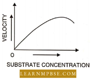
- The rate of enzyme reaction is directly proportional to the substrate concentration
- Presence of an enzyme inhibitor in the reaction mixture
- Formation of an enzyme-substrate complex
- At higher substrate concentration the pH increases.
Answer: 2. Presence of an enzyme inhibitor in the reaction mixture
Question 48. Which one of the following four secretions is correctly matched with its source, target and nature of action?
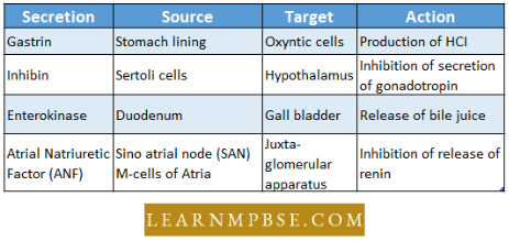
Answer: 1.
Question 49. Maltase converts,
- maltose to glucose at pH greater than 7
- maltose to glucose at a pH lesser than 7.0
- maltose to alcohol
- starch to maltose at pi 1 higher than 7.0,
Answer: 1. Maltose to glucose at pi I greater than 7
Question 50. Which pair is essential for the growth of first in water?
- calcium and phosphorus
- phosphates and carbonates
- sulphate and carbonates
- nitrates and sulphates.
Answer: 1. calcium and phosphorus
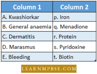
Question 51. Match the following nutritional vitamin deficiencies in column 1 with the causes/deficiencies in column 2 and choose the correct option from the answer key.
- A = p, B = t, C = q, D = r, E = s
- A = t, B = q, C = r, D = s, E = p
- A = t, B = r, C = s, D = p, E = q
- A = r, B = s, C = p, D = t, E = q.
Answer: 4. A = r, B = s, C = p, D = t, E = q
Question 52. The main function of lacteals in the villi of human small intestine is the absorption of:
- amino acids and glucose
- glucose and vitamins
- water and mineral salts
- fatty acids and glycerol.
Answer: 4. fatty acids and glycerol
Question 53. Which of the following is called a detritivore?
- an animal feeding on decaying organic matter
- an animal feeding on a plant
- a plant feeding on an animal
- an animal feeding on another animal.
Answer: 1. an animal feeding on decaying organic matter
Question 54. Which one of the following enzymes carries out the initial step in the digestion of milk in humans?
- Pepsin
- Rennin
- Lipase
- Trypsin.
Answer: 2. Rennin
Human Digestive System NEET Biology
Question 55. Examination of the blood of a person suspected of having anaemia shows large, immature, nucleated erythrocytes without haemoglobin. Supplementary his diet with which of the following, is likely to alleviate his symptoms?
- Riboflavin
- Iron compounds
- Thiamine
- Folic acid and cobalamine.
Answer: 4. Folic acid and cobalamine
Question 56. Which of the following pairs of the kind of cells and their secretion is correctly matched?
- oxyntic cells – a secretion with pH between 2.0 and 3.0
- alpha cells of islets of Langerhans – secretion that decreases blood sugar level
- Kupffer cells – a digestive enzyme that hydrolyses nucleic acids
- sebaceous glands a socialism trial evaporates on cooling,
Answer: 1. oxyntic cells – a secretion with pH between 2.0 and 3.0
Question 57. The following is a scheme showing the rate of carbohydrate timing digestion in the human alimentary canal. Identify the enzymes acting at stages indicated as a, b, c and d. Choose the convict option from those given
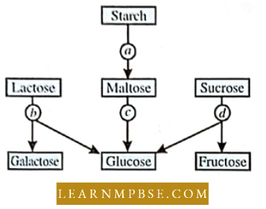
- a = amalyse, b = maltase, c = lactase, d = invertase
- a = amylase, b = maltase, c = invertase, d = lactase
- a = amylase, b = invertase, c = maltase, d = lactase
- a = amylase, b = lactase, c = maltase, d = invertase.
Answer: 4. a = amylase, b = lactase, c = maltase, d = invertase.
Question 58. Which one is correctly matched?
- vitamin E-thiamine
- vitamin D-riboflavin
- vitamin B1 -tocopherol
- vitamin A-calciferol
- vitamin B12-cyanocobalamine.
Answer: 5. vitamin B12-cyanocobalamine
Question 59. Which of the following is the correct chronological order for enzyme activity of some enzymes taking part in protein digestion?
- pepsin→ trypsin -4 peptidase
- pepsin →peptidase →trypsin
- trypsin → pepsin → peptidase
- peptidase → trypsin → pepsin
Answer: 1. pepsin →trypsin -4 peptidase
Question 60. Which of the following can be absorbed by hepatic caeca?
- glucose and amino acid
- glucose and lipid
- lipid
- glucose.
Answer: 4. glucose
Question 61. Which of the following hormones helps in the secretion of HCL from the stomach?
- renin
- gastrin
- secretin
- somatomedin,
Answer: 2. gastrin
Human Digestive System NEET Biology
Question 62. Statements
- The element which is very important for the production of thyroxine is iodine
- Vitamin B6 is otherwise known as niacin or nicotinic acid
- Fructose is a monosaccharide and is a hexose sugar
- Globulin is an example of a conjugated protein of the above statements.
- 1, 2 and 3 are correct but 4 is wrong
- 1 and 3 are correct but 2 and 4 are wrong
- 1 and 2 are correct but 3 and 4 are wrong
- 1 is correct while 2, 3 and 4 are wrong
- 1, 3 and 4 are correct but b is wrong.
Answer: 2. 1 and 3 are correct but 2 and 4 are wrong
Question 63. ‘Crypts of Liebcrkuhn’ are found in :
- gall bladder
- liver
- pancreas
- intestinal tracts
- salivary glands.
Answer: 4. salivary glands
Question 64. Glisson’s capsule is associated with
- liver
- pancreas
- lung
- Kidney.
Answer: 1. liver
Question 65. The main digestive function of enterokinase is :
- conversion of casein into paracasein
- conversion of pepsinogen into pepsin
- conversion of trypsinogen into trypsin
- Stimulation of the gastric glands to secrete gastric juice
- conversion of trypsin into trypsinogen.
Answer: 3. conversion of trypsinogen into trypsin
Question 66. Which of the following is the correct matching of the site of action on the given substrate, the enzyme acting upon it and the end product?
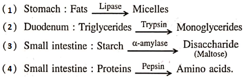
Answer: 3
Question 67. What will happen if the secretion of parietal cells of the gastric glands is blocked with an inhibitor?
- Gastric juice will be deficient in chymosin
- Gastric juice will be deficient in pepsinogen
- In the absence of HCL secretion, inactive pepsinogen is not converted into the active enzyme pepsin
- Enterokinase will not be released from the duodenal mucosa so trypsinogen is not converted to trypsin.
Answer: 3. In the absence of HCL secretion, inactive pepsinogen is not converted into the active enzyme pepsin
Question 68. Anxiety and eating spicy food together in an otherwise normal human may lead to:
- Jaundice
- Diarrhoea
- Vomiting
- indigestion.
Answer: 4. indigestion
Question 69. Select the correct match of the digested products in humans given in column 1 with their absorption site and mechanism in column 2. ‘
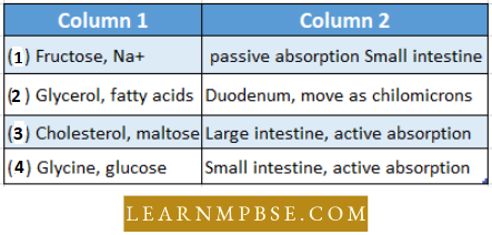
Answer: 4. Glycine, Glucose – Small Intestine, Active absorption