
Plant Anatomy (Meristem Plant Tissues Anatomy Of Root Stem And Leaf)
Meristem
Meristem. A meristematic tissue consists of a group of cells which have the power of division. The word meristem lias its origin from the Greek word ‘meristos’ which means divisible. (Nageli, 1058)
They comprise isodiametric immature cells that are undergoing division and expansion.
- They are densely organized to eliminate intercellular gaps.
- The cells are predominantly spherical and possess slender walls encasing them.
- Each cell comprises a conspicuous nucleus and thick protoplasm.
- Vacuoles in the cell are typically missing; when present, they are rather tiny.
- During cell division, the initial nucleus undergoes mitotic division into two nuclei, followed by cytokinesis, culminating in the production of two daughter cells.
- Proplastids are present in lieu of plastids.
- Metabolic activity is elevated.
- The cambium cells are elongated and contain vacuoles.
Read and Learn More NEET Biology Notes
Anatomy Of Flowering Plants Neet Notes
Anatomy Of Flowering Plants Neet Notes
1. Types Of Meristem Based On Development It is of three types :
- Promeristem
- Primary meristem
- Secondary meristem.
- Promeristem. It is made up of a group of meristematic cells that represent the youngest stage of a growing organ. It is the earliest stage of the primary meristem.
- Primary Meristem. Promeristem gives rise to primary meristem. By the rapid cell divisions, the cells of primary meristem get differentiated into different tissues. Cells of this meristem divide in three or more planes.
- Secondary Meristem. It develops at a later stage in the development of an organ. It is always lateral in position.
2. Meristematic Tissue-Based On Function: The first formed meristem (Promeristem) gets differentiated into the following three regions :
- Protoderm (gives rise to epidermis).
- Procambium (gives rise to phloem, xylem and cambium).
- Ground meristem (forms cortex, pericycle, medullary rays, pith, hypodermis and
endodermis).
3. Types Of Meristem Based On Location The Body. According to location in the body meristems are of three types:
- Apical meristems
- Intercalary meristems
- Lateral meristems.
- Apical Meristem. They are present at the apices of the stem, root and branches. These are responsible for increasing the length. Many cells form the apical meristems. However, in pteridophytes, one cell constitutes the meristem. It includes both pro-meristem and primary meristem.
- Intercalary Meristems: These are the parts of apical meristems, which get separated from the apex due to the development of permanent tissue in between. Intercalary meristems are found at the base of leaves above the nodes For Example. grasses, Equisetum, or below the nodes For Example. Mint. It increases the length of internodes and is finally consumed.
- Lateral Meristem. It is formed literally from the procambium strands i.e., cambium present between the xylem and phloem and the cork cambium of roots and stems. The cells divide only in the criminal plane and thus they add to girth. This type of meristem shows its activity after some time in plant organs.
4. Mcrislemulic Tissue-Based Oil Plane Of Cell Division:
- Mass Meristem. Here cells divide in all the planes thus increasing in volume. It can be noticed in the meristem of the cortex, pith etc.
- Plant Meristem. The cells divide in two planes. They form flat surfaces by increasing the area of organs For Example. Leaf formation.
- Rib Meristem. The cells divide only in one plane For Example., the formation of filaments in algae.
5. Based On Obesogens
- Dermatogen (forms piblcma and root cap in dicots),
- Periblem (forms cortex and endodermis), and
- Plcrome (forms pericyclic. vascular strand and pith.).
Anatomy of Flowering Plants – Important Notes for NEET Theories of Apical Meristem. Regarding the apical meristem, there are three theories:
- Apical Cell Theory Was Formulated By Nagcli (1858). According to the apical cell theory or Nagcli ( 1858), the apical meristem is made up of a single apical cell in all plants. This is also called “Single-cell theory”.
- Histogen Theory Proposed By Hanstein (1870). According to the history theory of Hanstein ( 1870), the apical meristem is divisible into three zones, i.e. outer dermatogen, middle problem and inner plenty. Dermatogen gives rise to epidermis, periblem to cortex and the pleronte to stele.
- Tunica Corpus Theory Was Formulated By Schmidt (1924). According to the tunica corpus theory of Schmidt (1924), the apical meristem is made up of two zones i.e. tunica and corpus. The tunica zone is outer in position and its cells divide only anticlinally. On the other hand, the corpus zone is central in position and its cells divide in many planes. Tunicacorpus theory is the most accepted theory of apical meristem.
- Korpe Kappe Theory (Schcupp, 1917). It deals with the organisation of the root apex, i.e. It has two parts. Kappe or cap and Korpe or body. Both show T-type division, upright or inverted.
Anatomy of Flowering Plants – Important Notes for NEET Quiescent Centre
Clowes (1961) has shown the occurrence of an inactive centre in many roots called a quiescent centre. It is the region of low mitotic activity which occupies the central region of the root apex, For Example., between the root tip and meristem. In the quiescent centre, there is little synthesis of protein DNA and RNA.
The root apex is differentiated into 3 regions-
- Protoderm (future epidermis),
- Cortical initials (future cortex) and
- Vascular cylinder. A group of initials at the apex produce cells on both sides.
Those towards the axis form the epidermis, cortex and vascular cylinder, while those away from the axis form the root cap. The Histogen involved in the formation of the cap (caiyptrogen) is derived from dermatogen.
Anatomy Of Flowering Plants Neet Notes
Neet Biology Anatomy Of Plants
They are arranged in the following four ways :
- Ranunculus Type. There is a single set or layer of initials that gives rise to all parts.
- Casuarina Type. Two sets or layers of initials-inner forming the central part, outer forming cortex, root cap and epiblema.
- Common Dicot Type. Three sets or layers of initials, one forming root cap and epiblema, second cortex and third central cylinder.
- Common Monocot Type. Four sets of initials, one (caiyptrogen) forming root cap, second epiblema, third cortex and fourth central cylinder.
Stem A place Meristem (Vetictatlve Shoot Apex)
It is a multicellular, dome-shaped or conical structure which is protected by young leaves produced by it. New leaves and axillary buds are produced periodically on the flanks.
- The period between the origin of two successive leaves is called plastoehron. The meristem forms three derivatives- protoderm (produces epidermis of stem and leaves).
- Procambium (forms vascular strand) and ground meristem (ground tissues like mesophyll of leaves, hypodermis, cortex. endodermis, pith, etc).
Anatomy of Flowering Plants – NEET Questions Permanent Tissue
Permanent Tissue. They are composed of mature cells which after undergoing complete growth, have assumed a definite shape, size and function. They have the power of division. Depending upon origin, permanent tissue is of two kinds:
- Primary tissue consists of evils derived from the primary meristem and
- Secondary tissue haring cells derived front secondary meristem.
The permanent tissue is of two types depending upon constitution i.e., simple tissue (one type of cells) and compound tissue (two or more than two types of cells.)
Kinds Of Simple Permanent Tissue.
- Parenchyma
- Collenchynta
- Sclerenchynta.
1. Parenchyma. Oval, spherical polygonal or isodiametric cells with thin cell walls. often with intercellular spaces.
- Parenchyma is modified into
- Chlorenehyma. These cells have chloroplasts and hence take part in photosynthesis. The Chlorenehyma of the leaf is called mesophyll. It is often differentiated into palisade parenchyma (columnar chlorenehymatous cells) and spongy parenchyma (irregular chlorenchyma cells enclosing air spaces),
- Aerenchyma. Network of star-like parenchyma cells enclosing large air cavities, For Example., aquatic plants.
- Epidermis. Cutinised parenchyma cells form a covering layer. A distinct layer of cutin or cuticle may occur on the outside,
- Guard Cells. They are a pair of specially thickened small reniform or dumb-bell cells which can create a pore in between them due to differential swelling,
- Prosenchyma. Elongated fibre-like parenchyma.
Anatomy Of Flowering Plants Mcq For Neet
- The tissue takes pan in the storage of food, slow conduction and turgidity of softer pans.
- It is the major ground tissue that occurs inside the stem and root as cortex and pith, the pulp of the fruit.
- The endosperm of the seed, parts of vascular tissues, leaf interior, etc. The epidermis is made of modified parenchyma cells.
2. Collenchyma. The tissue of cells with thick cellulose cell walls, especially at the angles of the cells is called collenchyma. It is abundant in climbing stems. It occurs in the hypodermis of dicot stems and petiole.
Depending upon the place of thickening, collenchyma is of three types:
- Lamellate (Lamellar). Thickening on tangential walls For Example., the stem of a Sunflower,
- Lacunate. Thickening on the walls bordering intercellular spaces, For Example.. stem of Tagetes. Tomato. Datura.
- Angular. Thickening at the angles. For Example.. stem of Cucurbita.
Flowering Plant Tissue Structure Neet
Anatomy of Flowering Plants – NEET Questions Collenchyma Functions.
- It provides mechanical support.
- It provides elasticity while allowing plant organs to grow in size.
- The cells may store food as well as manufacture food.
3. Sclerenchyma. It is made up of hard and lignified tissue consisting of fibres and sclereids. Its function is also supportive.
- There are two types of sclerenchyma cells
- Long, flexible with tapered ends (fibres) and
- Usually rough and spherical sclereids or stone cells.
Sclerenchymatous fibres. Individually, fibres are quite strong due to lignified walls. When these groups are together, their strength is greatly increased.
- Therefore, these can provide mechanical support to the tissue in which these are found. Fibres are commonly found in the pericycles of stems, forming a solid tissue protecting the vascular bundles of dicots.
- These also surround the vascular bundles of monocots and quite often, they form a layer in the cortex below the epidermis of stems or roots. These also occur in the xylem and phloem.
Anatomy of Flowering Plants – NEET Questions Sclerenchyma Commercial Fibres
- Surface fibres For Example. cotton, calotropis, and coir (coconut).
- Bast Fibres For Example. Hemp, Linum. Jute, Sun hemp.
- Leaf Fibres For Example. Agave, Murs.
Steroids are usually singly or in groups in the plant body, especially in the phloem, fruit walls and in the pulp of fruits and seeds, Sclereids provide rigidity to the structures in Which these are found.
The graininess of the fruits like guava and pears is due to the occurrence of stone cells in soups in their pulp. The ssalls of sclereids are very’ thick, and highly lignified so that lumen is greatly reduced.
Sclerites-Sclereids occurring singly are called idioblastic sclereids or spicular cells or sclerites.
Anatomy of Flowering Plants – NEET Questions Sclereids Are Of The Following Five Types
- Brachysdereids (Stone cells). Isodiametric sclereids resembling parenchyma in shape. found soft parts of plants.
- Macrosdereids (rod cells or Malpighian cells). Columnar b shape, forming palisade-like epidermal layer b seed coats of peas and beans and epidermis of onion.
- Osteosclereids (prop cells). Bone-like sclereids, columnar with dikted ends For Example. leaves and seed coat of many monocots. hr.
- Astrosclereids-star-like (stellate) sclereids For Example. Nymphaea leaves, stem and leases of Thea. (tea).
- Trichosclereids (or Trichoblasts or Internal hairs) are Long, hair-like, sometimes branched sclereids For Example. leaves of Olea and aerial roots of Monsiera.
Anatomy of Flowering Plants – NEET Questions Sclereids Functions
The function of sclerenchyma is to provide support and mechanical strength to the plant. Therefore, its distribution in plants is related to be physical stress which different organs have to face.
Sclereids Compound Tissue
Sclereids is two types, i,e. xylem and phloem.
Anatomy of Flowering Plants – NEET Questions Xylem
The xylem is a permanent complex conducting tissue. It is also called wood. It is concerned with the upward conduction of water and minerals.
Xylem is a group of cells which are similar b origin and function but of more than one type b structure. On be basis of development, the xylem consists of two primary xylem and a secondary xylem.
Primary Xylem. It is differentiated into :
- Protoxylem. It is the first formed part and consists of annular and spiral tracheids and vessels.
- Metaxylem. It is formed later and consists of scalariform, reticulate and pitted tracheids and vessels.
The Primary Xylem Is Of Three Types based on be relation between the protoxylem and metaxylem.
Three forms are:
- Exarch.
- Endarch
- Mesarch
The secondary xylem is formed from vascular cambium during secondary growth. It may consist of annual rings. Each annual ring is made up of autumn wood and spring wood.
Anatomy of Flowering Plants – NEET Questions Elements Of Xylem
Xylem is composed of the following four different kinds of elements:
- Tracheids
- Vessels
- Xylem parenchyma
- Xylem fibres.
1. Tracheids. These are elongated (1-6 mm) tube-like dead single cells with hard, thick and lignified walls. Their end walls are tapered and overlap with adjacent tracheids. When mature, babies are dead with empty lumens.
Neet Important Questions Anatomy Of Flowering Plants
Water can pass through empty lumens and passes from tracheid to tracheid through ‘pits’ in beer walls. However, angiosperms have relatively fewer tracheids, and vessels are more common b bese plants.
Anatomy of Flowering Plants – NEET Questions Depending Upon Thickenings, Tracheids Are Of The Following Type
- Annular,
- Spiral,
- Reticulate,
- Scalariform and
- Pitted (simple pits or bordered pits).
2. Vessels. These are characteristic conducting units of xylem m angiosperms. These are very long (l-6m) tubular structures formed by be fusion of several cells (called vessel elements) end to end in a row.
The vessel elements are similar to tracheids, except bats are shorter and wider than be latter. Vessels are exceptionally long in Eucalyptus.
3. Xylem Parenchyma. Parenchymatous cells associated with the xylem together constitute the xylem parenchyma. The cells are living, thin-walled and abundant. These are mainly involved in the short-distance transport of substances as well as the storage of sugars, starch and lipids.
4. Xylem Fibres. Sclerenchymatous fibres present in the xylem are called xylem fibres. These occur abundantly in woody dicots. These do not conduct water. Thus, these have thicker walls and narrower lumens than xylem vessels and are, therefore, stronger and provide additional mechanical strength to the xylem.
Anatomy of Flowering Plants – NEET Questions Phloem
It is a permanent complex tissue. It is meant for the conduction of food materials. Phloem occurs throughout the plant body along with the xylem. Phloem is of three types based on its position i.e. External phloem present outside the xylem. Internal phloem present inner to xylem and Interxylary or included phloem present within the secondary xylem.
Phloem is made up of four types of cells :
- Sieve tubes
- Companion cells
- Phloem parenchyma
- Phloem fibres.
1. Sieve Tubes. These are long, slender, tube-like structures involved in the transport of solution of organic solutes like sucrose throughout the plants.
- These are formed by end-to-end fusion of cells called sieve tube elements or sieve elements. Rows of these cells develop in the apical meristem together with the primary xylem.
- The walls of sieve tube elements are made up of cellulose and pectic substances, as in parenchyma cells, but their nuclei degenerate and are lost as they mature. The cytoplasm is confined to a thin peripheral layer.
- Two adjoining end walls of neighbouring sieve elements form a sieve plate. Originally plasmodesmata passed through the walls, but later on, these pores enlarged, so that the walls looked like a sieve allowing the flow of solution from one element to the next.
2. Companion Cells. A thin-walled elongated cell called a companion cell is associated with each sieve tube. Both are connected by simple pits.
Each companion cell is living and contains dense protoplasm and a large elongated nucleus. The sieve tube elements do not have a nucleus, but they remain living, being dependent upon the adjacent companion cell.
3. Phloem Parenchyma. These are parenchymatous cells found in the phloem. These are living and often cylindrical. These are absent in most of the monocots. These mainly store food materials.
4. Phloem Fibres. Phloem fibres are similar to sclerenchyma fibres and provide mechanical support. Phloem fibres of plants like jute, flax and hemp are retted in water and extracted for making ropes and coarse textiles. Sclereids are more frequent in older phloems.
Secondary Growth In Flowering Plants Neet – NEET Questions Types of Vascular Bundles
- Vascular Bundle. A strand-like part of the plant vascular system (conducting system) containing xylem and phloem. the xylem and phloem may be separated by the fascicular
cambium. the following types of vascular bundles are found in plants.
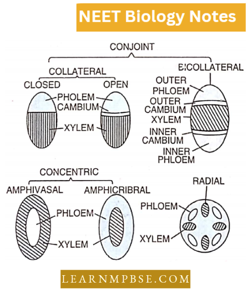
1. Conjoint Vascular Bundle. When phloem and xylem axes are present on the same radius, i.e. they are laterally placed to each other. It is of two types
- Collateral and
- Bicollateral.
- Collateral Vascular: If a strip of cambium is present between the phloem and xylem, they are called open bundles. If cambium is absent they are called closed bundles. If above the conjoint bundle, a sclerenchyma fibre is present the bundle is called fibrovascular bundle.
- Bicollateral Vascular Bundle. When in a collateral bundle, the problem is present both above and below the xylem bundles on the same radius. One group of phloem faces the pericycle and the other group faces the pith. This is seen in the Cucurbita stem and the cambium twice between both phloem groups lying above and below the xylem.
2. Radial Vascular Bundle. When the xylem and phloem lie on different radii alternating with each other, as in roots, the bundle is called radial.
Anatomy Of Dicot And Monocot Plants Neet
3. Concentric Vascular Bundle. When the xylem is surrounded by phloem or vice versa, the vascular bundle is called concentric.
If the xylem is surrounded by phloem it is called a concentric amphicribal vascular bundle (Hydrocentric) i.e. Ferns (Lycopodium, Selaginella). But when a phloem is surrounded by a xylem, it is called a concentric amphivasal vascular bundle (Leptocentric) Example. Dragon plant (Dracaena) and Dagger plant (Yucca).
Anatomy Of Flowering Plants Chapter 6 Special Tissues
They are commonly secretory or excretory tissue For Example. laticiferous tissue and glandular tissue.
1. Laciceferous Or Laticiferous Tissues Or Laticifers
It is concerned with the secretion of latex or an emulsion or oils, alkaloids, resins, proteins and sugar. It consists of thin-walled, elongated branched ducts. The laticifers are of two types
- Latex vesseLs
- Latex celLs.
These occur irregularly distributed in the mass of parenchymatous cells.
- Latex Cells are called simple laticifers or non-articulated laticifers which may be unbranched or may be branched profusely but do not fuse to form a network quite common in the members of families Apocynaceae, Asclepiadaceae, Euphorbiaceae and Moraceae (For Example. milkweed. Euphorbia, Ficus etc.).
- Latex Vessels (Articulated laticifers) are formed by the union of latex cells (hence called compound Laticifers also) and are quite common in the family Papaveraceae (poppy family), Caricaceae (Papaya family), Compositae, Musaceae (banana family)
2. Glandular Tissue. They are isolated secretory structures which may be unicellular or multicellular. Example
- Nectar-Secreting glands.
- Nectar (or honey) is a sugary substance secreted by the nectar gland or nectaries which may be floral (present in flower) or extra floral (on vegetative parts).
- Water-Secreting Structures (Water Stomata or Hydathodes) are present along the margins and apices of leaves. The exudation of water through these is called guttation.
- Osmophores. They secrete essential oil.
- Chalk Or Gland cells secrete salts For Example. Tamarix.
Simplified Notes On Anatomy Of Flowering Plants For Neet
Sachs (1975) distinguished three types of tissue systems in plants
- Epidermal tissue system,
- Ground tissue system,
- Vascular tissue system.
1. Epidermal Tissue System
- It consists of the epidermis, the outermost layer of cells covering the entire surface of the plant body and epidermal outgrowths.
- The Epidermis of the stem, leaves and floral parts originate from the surface layer of the shoot apical cistern.
- In roots, the epidermis originates either from an independent Set of Initials or has a common origin with the root cap and cortex. It is called epithelia or piliferous layer, it may bear root hair.
In most of the angiosperms, it is single-layered (uniseriate), but in some (like leaves of Nerlum, Ficus, Olearulcr etc.) it is made up of two or more layers (multiseriate).
- Epidermal cells are living, thin-walled, with a large central vacuole and thin peripheral cytoplasm. These are compactly arranged without intercellular spaces. The basic function of the epidermis is to protect the plant from desiccation and infection.
- The epidermal cells secrete a waxy substance called cutin, which forms a layer of variable thickness (the cuticle) within and on the outer surface of its all walls. It helps in reducing the loss of water by evaporation and also checks the entry of pathogens.
- In Xerophytes, the cuticle is thick; in mesophytes, it is moderately thick; and is absent in hydrophytes. It is also absent in roots and underground parts.
Epidermal outgrowths are called trichomes (hairs or scales). The hair-like extensions may be unicellular or multicellular and serve a variety of functions.
In roots, Unicellular hairs are called root hairs, these absorb water and mineral salts. In stems, these are multicellular. Trichomes are epidermal outgrowths. Scales are flattened epidermal outgrowths.
Stomata occur in the epidermis of aerial parts. Depending upon the distribution of stomata, the leaves are
- Apple-Mulberry Type. Hypostomatic stomata only on the lower surface.
- Potato Type. Most common, amphistomatic but more on the lower surfaces,
- Oat Type. Amphistomatic, equal on two surfaces,
- Nymphaea Type. Epistomatic—only on the upper surface,
- Potamogeton Type. Astomatic or with nonfunctional stomata.
2. Ground Tissue System:
It comprises the internal structure of organs, excluding the circulatory system. The ground tissue system in leaves is referred to as mesophyll. It is categorized into hypodermis, cortex, endodermis, pericycle, pith, and medullary rays.
- The hypodermis imparts strength. The cortex is involved in food storage. In certain stems, the cells possess chloroplasts and are also photosynthetic.
- The cortex of immature roots transports absorbed water and minerals inward.
- The endodermis, the innermost layer of the cortex, contains Casparian strips in roots.
- In dicot stems, it is referred to as a starch sheath.
- The endodermis is lacking in the stems of monocots. The pericycle constitutes the external boundary of vascular tissues. It is unilayered in roots and multilayered in stems.
3. Vascular Tissue System:
It consists of a vascular strand or cylinder. The latter is made up of some vascular bundles. Vascular bundles are radial in roots and conjoint in other parts.
Plant Tissues And Anatomy Neet Questions
Anatomy Of Flowering Plants Chapter 6 Anatomy Of Root
Internal Structure of Dicot Root
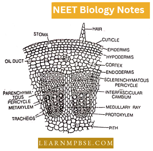
A primary root arises from the radicle of the seed. It is generally cylindrical in outline and possesses the following structures, from outside towards the inside of the transverse section of the root:
- Epiblema Or Piliferous Layer. It is the outermost layer which is made up of compactly arranged thin-walled parenchymatous cells. From epidermal cells, arise thin-walled tubular outgrowths called root hair. Due to the presence of root hair, the epiblema layer is also called as piliferous layer. The Epiblema layer is responsible for the absorption of water and minerals from the soil.
- Cortex. Inner to epiblema is a multilayered cortex which is made up of thin walled cells. The cells of the cortex store food. These cells also conduct water from epiblema to internal tissue.
- Endodermis. The innermost layer of the cortex is known as the endodermis. The endodermis consists of barrel-shaped cells with Casparian strips on their anti-clinal walls. Opposite to the protoxylem, the cells are thin-walled and called passage cells. Passage cells allow the free movement of water and minerals from the cortical cells in the xylem bundles.
- Pericycle. Inner to endodermis is a layer of pericycle. From the pericycle arise lateral roots and also vascular cambium which brings about secondary growth in the root.
- Vascular Bundle. Inner to pericycle are found 2-6 vascular bundles with alternating xylem and phloem. According to number, the root may be diarch, triarch, tetrarch and pentarch.
In the xylem, the protoxylem is outside and the metaxylem is towards the centre of the root. Such a xylem is called an exarch.
- Phloem is present in between two xylem bundles. Phloem consists of sieve tubes, companion cells and phloem parenchyma.
- Dicot root shows secondary growth. Cambium develops from pericycle and conjunctive parenchyma, and a ring of cambium is formed which cuts off the secondary xylem on the inner side and the secondary phloem on the outer side.
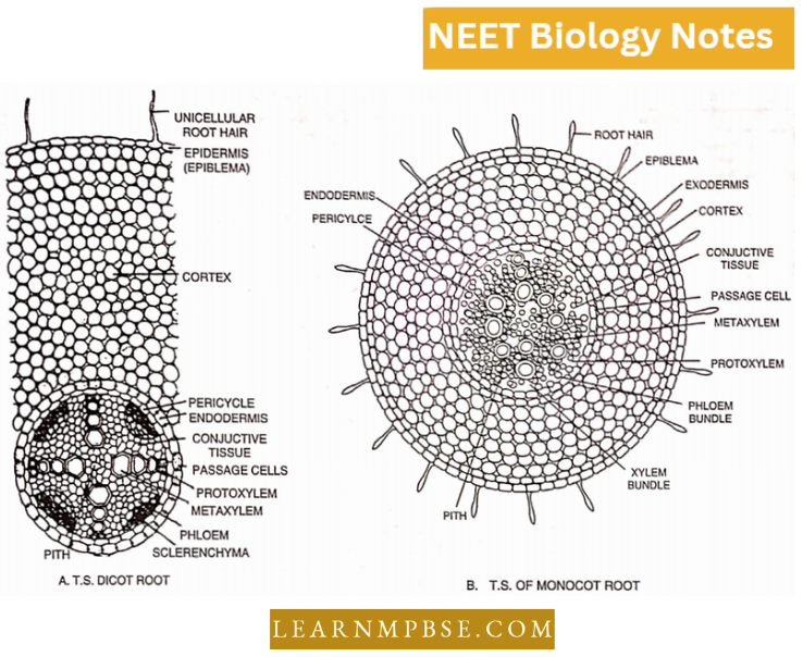
Anatomy Of Flowering Plants Chapter 6 Internal Structure of Monocotyledonous Root
The transverse section of the monocot root shows the following structures. There is no distinction between young and old monocot roots as there is no secondary growth in monocot rool The various layers of tissues are as follows:
- Epiblema Or Piliferous Layer. It is the outermost layer which consists of thin-walled cells. Some cells produce root hair. It is meant for the absorption of water.
- Cortex. Inner to epiblema is a multilayered cortex consisting of compactly arranged cells. It is meant for protection and storage of food.
- Endodermis. It is single-layered and lies inner to cortex. The cells are barrel-shaped with Casparian strips on their radial walls. Opposite to protoxylem, endodermal cells are thin-walled and called passage cells.
- Vascular Strand. It consists of several (8 or more) alternate radial xylem and phloem bundles. The vascular bundles are arranged in the form of a ring with a pith in the centre.
- The xylem Is Exarch with a protoxylem towards the outside and a metaxylem towards the centre. Xylem elements are rounded. The xylem provides mechanical strength and is meant for the conduction of water and minerals.
- The Phloem Alternates With The Xylem. The two are separated by conjunctive parenchyma. Phloem is meant for translocation of organic food.
- Pith. Pith is present in the centre, consisting of parenchymatous cells. Pith stores food.
Anatomy Of Flowering Plants Chapter 6 Anatomy Of Stem
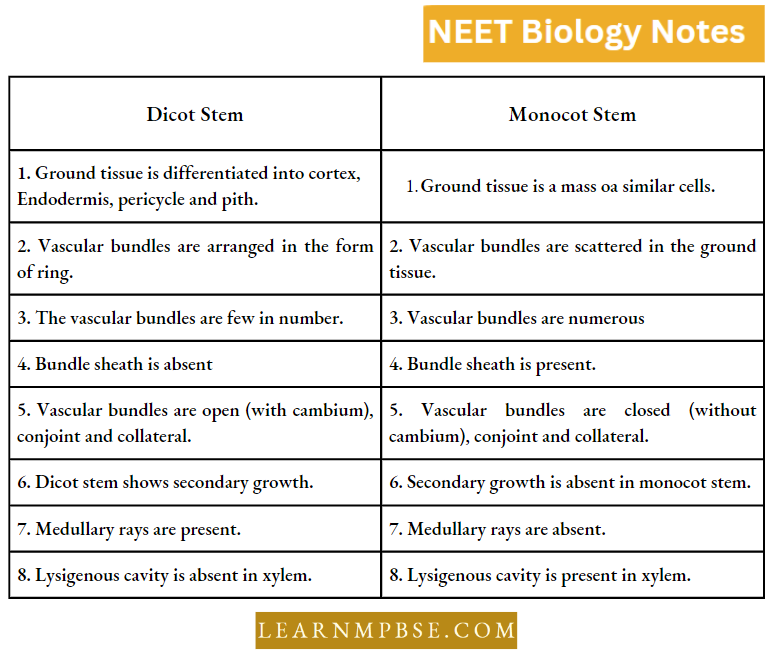
Structure Of Dicot Stem
A young dicot stem in the transverse section shows the following structures from the periphery towards the centre.
- Epidermis
- Cortex
- Endodermis
- Pericycle
- Vascular bundles
- Pith.
Structure Of Monocot Stem
In the monocot stem, there is no distinction between the cortex and the pith. There is ground tissue in which vascular bundles are scattered. The transverse section of Maize stems shows the following structures:
- Epidermis
- Ground tissue.
- Vascular bundles.

Anatomy Of Flowering Plants Chapter 6 Anatomy Of Leaf
Internal Structure Of A Dicotyledonous Leaf Or Dorsiventral Leaf of Helianthus or Chenopodium can be studied under the following heads :
1. Epidermis. In the transverse section, there are upper epidermis and lower epidermis. The epidermis is single-layered. The cells of the epidermis are colourless and secrete a waxy layer of cuticle.
- In the epidermis are numerous minute apertures called stomata. Each stoma has two kidney-shaped guard cells. Stoma opens in the leaf in a sub-stomatal cavity.
- Besides guard cells, there are two specialised cells called subsidiary cells or accessory cells. The guard cells regulate the opening of a stoma.
Plant Tissues And Anatomy Neet Questions
2. Mesophyll. In between the upper and lower epidermis, there is chloroplast containing photosynthetic tissue called mesophyll. Mesophyll is divided into two parts:
- Palisade parenchyma
- Spongy parenchyma.
- Palisade Parenchyma. It is present below the upper epidermis and consists of closely packed elongated cells. The cells contain abundant chloroplasts and are arranged in 2-3 layers.
The compactness of the cells of this layer reduces the transpiration. The main function of palisade tissue is to manufacture carbohydrates during photosynthesis.
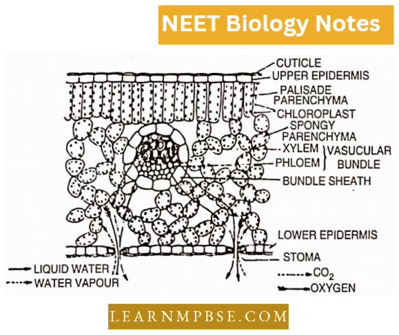
- Spongy Parenchyma. It is present below palisade tissue and consists of loosely arranged irregularly shaped cells with large intercellular spaces in between them.
Spongy parenchyma is in contact with the atmosphere through stomata. Its function is transpiration and exchange of gases for respiration and photosynthesis. In addition, spongy parenchyma is photosynthetic in function.
3. Vascular System. There are many vascular bundles. Each vascular bundle is surrounded by a layer of parenchymatous cells called a bundle sheath. Each vascular bundle has a xylem towards the upper epidermis and a phloem towards the lower epidermis.
The xylem is meant for the conduction of water and minerals while the phloem is meant for the translocation of food. Cambium is absent in the leaf, so there is no secondary growth in the leaf.
Structure of A Monocotyledonous Leaf Or Isobilateral Leaf Grass Or Zea Mays.
The transverse section of a monocot leaf shows the following parts:
- Epidermis. It consists of a closely packed single layer of cells. Some cells in the epidermis are large, and thin and contain water and are called bulliform cells. In epidermis are present stomata. Each stoma has two dumbbell-shaped guard cells.
- Mesophyll. There is no distinction between palisade and spongy parenchyma. The mesophyll consists of similar types of cells rich in chloroplast. There is no distinction between palisade and, spongy parenchyma. The main function of mesophyll is photosynthesis.
- Vascular System. The vascular bundles are present in a row. Each vascular bundle is surrounded by a sclerenchymatous bundle sheath. In the vascular bundle xylem is towards the upper epidermis and the phloem towards the lower epidermis. There is no cambium. So vascular bundle is closed.
Secondary Growth
Secondary Growth is growth brought about by secondary meristem. It may be classified into two types:
Normal And Anomalous. Growth brought about by fascicular cambium is called normal. Secondary growth of vasculature brought about by extra fascicular cambium (arising either in the pith or cortex) is called anomalous.
- In dicots, secondary growth is seen in the vascular cylinder as well as in the cortex. Vascular cambium produces secondary vascular tissue while cork cambium produces periderm tissue.
- In monocots vascular bundles are closed, thus there is no secondary growth. In some monocots, Dracaena, Yucca, Aloe, Agava, and Sanserveria, secondary growth occurs.
Anatomy Of Flowering Plants Chapter 6 Steps Of Secondary Growth
- Secondary growth results from the activities of the cambium and cork cambium.
The cambium is a lateral meristem that is one cell thick radially. - It consists of fusiform initials and ray initials. It may be multi-storeyed or single-storeyed.
- A cambium ring consisting of intra- and interfascicular cambial strips is established.
- It generates secondary xylem internally and secondary phloem externally. Additionally, secondary medullary rays are produced on both sides.
- The secondary xylem and phloem form a vertical and a horizontal system. The latter in both instances consists of ray parenchyma.
- The secondary medullary rays consist of upright and reclining cells. The rays might be either homocellular or heterocellular. They can be homogeneous or heterogeneous, and uniserate, biserate, or multiserate.
- The growth throughout the spring and fall seasons results in spring and autumn woodlands, respectively. The two types of wood form the annual ring. The age of the plant can be approximated by counting the annual rings.
- The wood can be either porous or non-porous, with the porous category further classified as ring porous or diffuse porous.
- The paratracheal parenchyma generates balloon-like formations known as tyloses within the tracheary components.
The wood is thereafter categorized into a narrow, light-hued region known as sapwood or alburnum, and a dark-hued, large region referred to as heartwood or duramen.
- The sapwood is physiologically active, facilitating the ascent of sap. The heartwood has more resistance than sapwood. Reaction wood develops as a response to stress. It could be either tension wood or compression wood.
- The extra-stellar zone expands when a cork cambium (phellogen) develops in the cortex or epidermis.
- The phellogen generates cells outside that undergo suberization to produce cork (phellem). The cells on the inner side remain thin-walled and constitute the secondary cortex (phelloderm).
- The three layers, namely phellem, phellogen, and phelloderm, collectively form the periderm. Non-suberized cells of phellem are referred to as spheroids.
- All tissue external to the vascular cambium is referred to as bark. The tissue external to the innermost phellogen forms the outer bark or rhytidome, while the remaining tissue is classified as the inner bark.
- The bark can be scaly, papery, ring-shaped, or exhibit intermediate characteristics.
- Commercial cork is derived from Quercus suber. The initial layer is referred to as a virgin cork. It is lightweight, robust, flexible, a poor conductor of heat and electricity, and impermeable to liquids and gasses.
- A wound, once created, is promptly filled with cork cells known as wound cork, produced by the activity of phellogen.
- A lenticel develops at the stomatal site. The sub-lenticular cells serve as the complimentary cells. A phellogen is established, generating new complimentary cells and protective layers.
- When branches accelerate in the stem as a result of secondary growth, nodes are generated.
Anatomy Of Flowering Plants Chapter 6
Plant tissue may be classified into two main groups i.e. meristematic tissue and permanent tissues.
- According to position, the meristem is of three types i.e. apical, intercalary and lateral.
- The apical meristem lies at the apex of both stem and root.
- Various types of trichomes are
- Simple
- Unicellular,
- Multicellular
- Multicellular with protuberance and
- Multicellular
- Branched and
- Stinging and
- Glandular
- In a dicot root, there are 2 to 6 xylem bundles while in monocots there are more than six.
- Latex of Carica papaya (papaya) contains papain. Latex of poppy yields opium which contains the alkaloid morphine.
- Latex of Bananas contains tannin.
- Though Hevea brasiliensis (Para rubber) and Manihot glaziovi belong to the family Euphorbiaceae, these contain latex vessels (not latex cells).
- Latex cells are found in Cannabis, Vinca minor and Urtica dioica. Para rubber and Indian rubber are obtained from the latex of Hevea brasiliensis and Ficus elasticatively.
- Aristolochia And Bougainvillea show anomalous secondary growth.
- The periderm is a secondary protective tissue that replaces the epidermis during secondary growth. It consists of phellogen (cork cambium) which produces phellem towards the outside and phelloderm towards the inside.
- Endodermoid. The term is used by some authors for endodermis or starch sheath of young stems because of the absence of Casparian strips.
- Leaf Primordium. Develops from a lateral protrusion or leaf buttress. It grows initially by an apical meristem (permanent in ferns) and then by intercalary meristem.
- The leaf consists of only primary tissues. Secondary growth is limited to wound healing.
- The epidermis which covers the upper surface of the leaf is called the adaxial epidermis while that which covers the lower surface is known as the abaxial epidermis.
- In grasses and Equisetum, silica is present in the epidermal cells.
- Epidermal cells containing cystoliths are called lithocysts.
- The epidermis of garlic (Allium sativum), scales and seeds of peas and beans are made up of sclereids.
- Usually, epidermal cells are colourless, but hydrophytes and sciophytes, contain chloroplasts and are hence, green.
- Cork is light, highly compressible and does not catch fire.
- In the deserts; 60 cm. high plant Aerva persica plant can have a tap root system reaching a depth of 6 metres.
- Apoplast. The non-living parts (For Example. xylem, cellulose, intercellular space, etc.) of the plant are called apoplast.
- Symplast. Living parts (cytoplasm-containing cells) of the plant.
- Sclerenchymatous patches of the pericycle outside the vascular bundles are called bundle caps.
- Lateral roots arise endogenously from the pericycle cells.
- Medullary vascular bundles are found in the stems of Mirabilis, Boerhaavia, Amaranthus, etc. The plants show anomalous secondary growth.
- Abscission. It involves the formation of a special parenchymatous layer called abscission or separation layer at the base of the organ and a layer of suberised thick-walled ‘cork’ cells called a protective layer over the mother axis. Degeneration of cells of abscission or separation layer causes abscission.
Anatomy Of Flowering Plants Chapter 6
Vessels are an advanced type of conducting element and are characteristically found in angiosperms. Some primitive vessel-less angiosperms belong to the families—Winteracae, Trochodendraceae and Telracentraceae. Some pteridophytes (Selaginella, Pteridium) and gymnosperms (Gnetum) have got vessels.
- Wood without vessels is homozygous while the one with vessels is heterozygous.
- Gymnosperms With Vessels. Members of group gnetales.
- Pteridophytes With Vessels. Occasional in species of Selaginella, Dryopteris, Marsilea, etc.
Fibres present outside the xylem are called extrasolar fibres. They may be cortical, pericyclic (or perivascular) or phloem (bast) fibres.
- Except xylem parenchyma, the xylem is a dead tissue.
- Sieve tubes were first discovered by Hartig (1837).
- In phloem, companion cells and sieve tubes arise from the same mother cell.
- Slime plugs are dense funnel-shaped structures formed by the coagulation of slime bodies on sieve plates.
- conifers (gymnosperms), albuminous cells are found-analogous to companion cells.
Since companion cells and sieve tubes arise from the same mother cell, these are called sister cells.
- Knot. As the stem grows in thickness, the bases of branches become embedded in the secondary xylem and thus knots are formed. The buried portion can neither grow in diameter nor can be pushed outward.
- When a log is cut vertically, the branch embedded in it as a knot, is cut transversely.
- P-proteins are proteinaceous structures present in sieve tubes and are believed to be responsible for
- Movement of materials through the cell,
- Sealing of pores after wounding.
- Bhojpatra is derived from the bark of Betula utilis.
The waxy substance associated with the walls of cork cells is suberin and the phenomenon of impregnation of cell walls with suberin is called suberisation.
- Cutin forms a continuous layer on the epidermis, which is known as a cuticle.
- The formation of the cuticle is called cuticularization.
- Apical cell theory was proposed by Hofnieister (1857) and supported by Nageli.
- Histogen theory was given by Hanstein (1868) and supported by Strassburger.
- Heartwood. Most abundant in Mulberry but absent in Poplar and Willow.
- There is no distinction between heart wood and sap wood in Salix, Populus, etc.
- In Morus, Taxus, the heartwood is most abundant and the sapwood zone is quite thin.
Heartwood is dark-coloured due to the deposition of extractives and is considered durable.
- Heartwood is the dead primary or old xylem. Most Durable Soft Wood. Cedras deodara.
- Most Durable Wood. Teak (Tectona grandis).
- Lightest Wood. Ochroma pyramidale ( = 0. lagopus).
- Heaviest Wood. Guaiacwn officinale. In India Acasia sundra.
- The bark of Cinnamomum leylanicum (Dalchini) is used as a flavouring material.
- In Angiosperms vessels are present along with tracheids.
- In the sieve tube nucleus is absent.
Neet Important Questions Anatomy Of Flowering Plants
Companion cells occur only in Angiosperms and are absent in Pteridophytes and Gymnosperms.
- Phloem parenchyma is absent in monocots.
- Xylem cells are polygonal in dicots and oval in monocots.
- Normally no secondary growth in monocot stems except anomalous secondary growth in Dracaena, Yucca etc.
- In monocots, grafting is not successful due to closed and scattered vascular bundles.
- In a hollow-hearted plant, the growth of the plant is not affected.
- Wound healing and secondary growth are controlled by secondary meristems.
Swollen exogenous protuberances on the growing apices are leaf primordia.
- Sieve elements are living enucleated structures.
- Tissue builder zones are three in the shoot apex and four in the root apex.
- Raphides are the crystals of calcium oxalate.
- Amphivasal is where the xylem surrounds the phloem.
- Clayptrogen histogen forms a root cap.
Climatic variations are almost zero in seashore plants therefore cambium has uniform activity without demarcation of annual rings.
- The leaf consists of only primary tissues. Secondary growth is limited to wound healing.
- Leaf primordium develops from a lateral protrusion or leaf buttress. It grows initially as apical meristem (permanent in ferns) and then by intercalary meristem.
- The epidermis which covers the upper side of the leaf is called the adaxial epidermis.
- The epidermis which covers the lower surface is called the abaxial epidermis.
- Cells of bark i.e. cork of phellem cells are made up of impervious suberin.
- After the formation of phellem, the stomata are obliterated and new proes are formed which are lens-shaped hence lenticels.