
NEET Biology Pteridophyta
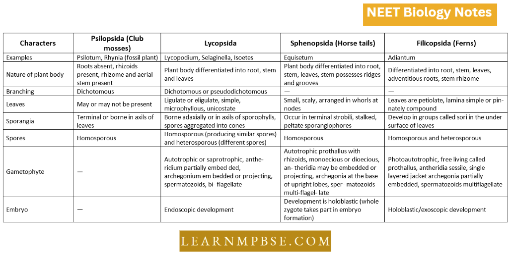
- Pteridophytes. The term Pteridophyta was coined by Haeckel. Pteron (Gk) means a feather and the name Pteridophyta was given to this group because of their pinnate or feather-like fronds. Pteridophytes include about 10,000 species.
- Ferns are a big group of pteridophyta with 9300 species and some ferns may be 18 metres in height (Giant tree fern).
- The smallest pteridophyte is Azolla (aquatic fern)
Read and Learn More NEET Biology Notes
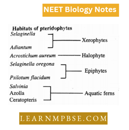
Habitats of pteridophytes
Pteridophyta NEET Notes
Features of pteridophytes
- Pteridophytes are assemblages of seedless vascular plants that have successfully invaded the land and reproduce through spores.
- Main plant body is sporophyte (2N).
- initiated into root, rhizome, and leaves. The latter may be megaphyllous or microphyllous.
- Plants show evolution of stele from protostele siphonostelesolenostele dictyostele dictyostele.
- Spores are produced in sporangia after meiosis. A group of sporangia occurs in the form of sorus covered with indusium. They may be homosporous (For example ferns) or heterosporous (For example Selaginella).
- Gametophytic Generation. Spores on germination form prothallus which is monoecious, bear both antheridia and archegonia.
- Sperms and eggs on fertilization form oospores which again start sporophytic generation
- The life cycle of Pteridophytes is of Diplohaplontic type i.e. main plant body is sporophyte which alternates with gametophyte. Both generations are morphologically dissimilar. This is also called a heterologous type of alternation of generations.
NEET Biology Pteridophyta Selaginella
The sporophytic generation of this lycopod has a plant body differentiated into genuine roots, stems, and leaves. The branched stem is typically horizontal and prostrate.
- Rhizophores extend vertically downward from the branches of the dichotomies. Adventitious roots form when the rhizosphere contacts the soil. The leaves may be uniform or potentially dimorphic.
- They possess microphyllous and ligulate characteristics. The sporophyte generates spores. Selaginella exhibits heterospory.
- Spores are generated in sporangia located in the axil of the sporophylls. The sporophylls are clustered at the terminal sections of the branches, forming the strobili.
- Each strobilus possesses microsporophylls and megasporophylls that are analogous and ligulate.
- The microsporophylls support the microsporangia, while the megasporophylls support the megasporangia.
- Each microsporangium and megasporangium possesses a bilayered jacket surrounding a nutritive tissue, the tapetum, which encases the spore mother cells. The majority of microspore mother cells undergo meiosis to produce many microspores.
- In the megasporangium, typically only one megaspore mother cell remains functional (sometimes 2-3), and after meiosis, it creates four megaspores, of which commonly one to three are nonfunctional. In S. motorsport, only a single megaspore is operational.
Plant Kingdom Pteridophyta NEET
- The developing spores are provided with nourishment by the remaining megaspore mother cells that endure degeneration
- The megasporangium assumes a lobed aspect as a consequence of the megaspores’ expansion. The spores are expelled through the dehiscence of the wall, which is divided into two halves by a vertical apical incision.
- The gametophytes. The spores are the initial cells of the gametophytic generation. The gametophyte’s growth is precocious and begins in the sporangia.
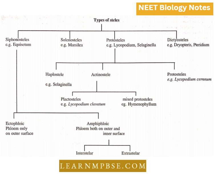
Modifications of siphonostctc
- Cladosiphonic siphonostele. A siphonostele not perforated by leaf gaps Example a few species of Selaginella.
- Phyllosiphonic siphonostele. A siphonostele perforated by leaf gaps- Example Nephrolepis.
- Solenostele. A siphonostele perforated by leaf gaps which are scattered but not overlapping Example Ferns.
- Dictyostele. A siphonostele perforated by several overlapping leaf gaps. Each separate strand is called a meri stele Example Dryopteris, Pteridium, Pteris, etc.
- Polycyclic dictyostele. A dictyostele consisting of two or more concentric rings of meristeles Example Pteridium aquilinum.
- Eustele. Much dissected siphonostele having vascular strands separated apart by parenchyma Example Equisetum.
- Polysetelic condition. Presence of more than one stele Example Selaginella kraussiana.
The microspores are orange-red, tetrahedral, with a two-layered wall and tri-radiate.
Their nucleus divides by mitosis to form male gametophytes within their wall. Their liberation is generally withheld upto the 13-celled stage.
At this stage, they contain 1 urothelial cell, 8 jacket cells, and 4 androgenic cells. The four androgenic cells divide to form the antherozoid mother cells and astrocytes and ultimately 256 antherozoids are formed.
Each antherozoid is biciliate and liberated by the rupturing of the exine. They swim about freely in search of a female gametophyte or if developed within the sporangium they may be carried along with the raindrops.
The megaspores are large, white to pale green, tetrahedral, and tri-radiate.
They may be shed before any trace of a cellular organization or shortly after the first archegonia are formed or may be retained until after fertilization and a considerable development of the embryo.
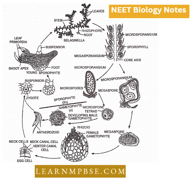
The germinating megaspore enlarges considerably followed by free-nuclear division eventually giving rise to a massive multicellular megagametophyte or female gametophyte tissue. A part of the tissue of the gametophyte gets exposed.
The exposed region may contain chlorophyll and even rhizoids. Several archegonia develop from the superficial cells of this tissue along with clusters of rhizoids.
The mature archegonia have a neck of two tiers of cells each, enclosing a neck canal cell and venter enclosing a ventral canal cell and an egg. At maturity, the neck and venter canal cells disintegrate exposing the egg to be fertilized.
Characteristics Of Pteridophyta NEET
Fertilization of the egg by the antherozoids results in the formation of the zygote or oospore.
Apogamy and parthenogenesis are known in several species.
The young Sporophyte. Following fertilization, the resulting zygote secretes a wall and divides and redivides to form the embryo. The embryo pushes its way into the tissues of the gametophyte by its suspensor.
It is differentiated into a stem apex, two leaf primordia with their ligules at one end, and the rhizosphere at the opposite end. Situated between them is the foot. This juvenile sporophyte ultimately matures into a sporophyte of Selaginella.
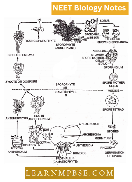
NEET Biology Pteridophyta Life Cycle Of Dryopteris
It belongs to the division Pteropsida, Class Filicineae, order Filicales, and family Polypodiaceae.
The plant body is sporophytic and the mature sporophyte contains roots, rhizomes, and leaves.
Roots are adventitious. The rhizome is well-branched and remains covered with several fine hairs called ramenta. Leaves are circinately coiled, when young. Mature leaves are very large and bipinnately compound.
Internally, the roots are ditch and exarch.
- Internally, the rhizome contains a dictyostele. Each dictyostele is made up of meristeles. Companion cells are absent in the phloem and the xylem is devoid of vessels.
- The stele is horseshoe-shaped in the upper portion of the rachis, while in its lower portion, it is made up of several meristeles.
- Spores are present in the sporangium. Several sporangia are grouped in a sorus. Several sori remain attached on the lower surface ofthe leaves, and such leaves are called sporophylls. Sori kidney-shaped bodies. Each sorus remains covered by a layer of indusium.
- Each sporangium is a stalked body. The edge contains 16-30, small brown cells called annulus. They possess thickenings on the inner and radial walls. The outer walls remain thin.
- A strip of thin-walled cells on each sporangium represents the stomium. The diploid spore mother cells, present inside the sporangium, divide by meiosis, and each forms four haploid spores.
- The spore germinates to produce a heart-shaped prothallus or gametophyte. Both the sex organs (antheridia and archegonia) and the rhizoids are present on the ventral surface of the prothallus. Archegonia are present in apical cushion and antheridia are present in rhizoidal region.
- Antheridia are present on the lower part of the ventral surface of the prothallus, along with rhizoids. Antherozoids are uninucleate and multiflagellate.
- Archegonia are present near the apical notch.
- Fertilization takes place in the presence of water. Malic acid is the chemotactic substance present in the archegonium. The zygote develops into a sporophyte without reduction division. The young sporophyte remains attached to the gametophyte for some time.
- Plants show heteromorphic alternation of generations.
Economic Importance
- Food. Sporocarps of Marsilea are edible. The starchy pith of Angiopteris erecta and Alsophila australis is eaten by the natives of Australia.
- Medicines. Rhizomes and petioles of Dryopteris have vermifuge properties. Adiantum roots can cure throat infections.
- Its leaves are dried and used as a laxative. Lycopodium is used in the treatment of rheumatism and disorders of the lungs and kidneys. Equisetum arvense is a diuretic while its ash relieves acidity and dyspepsia.
- Scrubbing. Equisetum stems have rough and abrasive surfaces. They are used in scrubbing and polishing.
- Ornamental value. Some ferns and club mosses are grown indoors as well as in gardens for their graceful foliage.
- A few characteristics that are responsible for the successful survival of vascular plants are: deep soil penetrating root system.
- Water-saving methods developed by plants as cuticles on leaves.
- support to woody plants is provided by collenchyma, sclerenchyma, and by the lignification of wood.
- they bear a vertical main axis and a conspicuous radial symmetry around the axis. Such construction permits efficient nutrient absorption from all sides around the plant. The pteridophytes were the first plants to develop vascular tissue.
- Pteridophytes are vascular plants without flowers and seeds.
- In pteridophytes sporophyte is independent and in bryophytes the sporophyte is dependent.
- The pteridophytes are the most primitive living (For example Selaginella, Lycopodium, Equisetum) and fossil (For example Rhynia) Vascular plants.
- The absence of root hair and root cap is a stem-like character of Selaginella rhizophore.
- They are regarded as organogenesis. Resembles with stem, exogenous origin, adventitious roots, and bear leaves when injured.
- Calixylon is a fossil pteridophyte from the upper Devonian period with a trunk of 5ft. These heterosporous pteridophytes must have given rise to gymnosperms.
- Simple sori (primitive) in which all sporangia develop simultaneously.
- Gradate sori (more advanced): where sporangia develop in basipetal succession or mixed sori (most advanced): where sporangia develop in an irregular sequence.
- Dryopteris and moss form monoecious gametophytes as all spores are uniform.
- Suigenesis means neither a root nor a shoot complete but something new.
- Circinate ptyxis is shown by young leaves that are under the influence of hyponasty and they take two years to open up under the influence of epinasty.
Examples Of Pteridophyta NEET
NEET Biology Pteridophyta Quanta To Memory
- Haeckel coined the term pteridophyta (vascular cryptogam).
- Zimmaman (1930) proposed the “Telome theory” for the evolution of vascular plants. He proposed that all vascular plants have evolved from simple dichotomously branched pteridophytes such as Rhynia.
- Tree fern with upright aerial stem Example Cyathaea, Alsophila.
- Cyathaea is the tallest fem
- Rhynia is a fossil plant placed under Psilophytales.
- Salvinia , Azolla and Marsilea are water ferns.
- Spores are usually all alike ( i.e. Homosporous) Example Lycopodium, Pteris, Adiantum, Pteridium, etc. or they are of two different sizes (i.e., Heterosporous) Example Selaginella, isolates, Marsilea, Azolla, Salvinia etc.
- Dryopteris is homosporous and Selaginella is heterosporous
- Sporangia are usually borne on or in the axil of leaves
called sporophylls. Examples are Lycopodium, Selaginella, and Pteris.
Nephrolepis, Adiantum, etc. In some aquatic plants
sporangia are produced within specialized structures called
sporocarps, for Example, Marsilea, Salvinia, Azolla, Regnellidium,
Pilularia etc. - In pteridophytes or Ferns, the archegonia secrete malic acid which attracts the antherozoids.
- In pteridophytes ovules are absent. Selaginella and fem both are pteridophytes, so in both ovules are absent.
- Azolla, a water fem is used as a biofertilizer.
- Ferns are cultivated as ornamental plants.
- The embryo is diploid and spores are haploid.
- In the fem, the neck is small and is having a single neck canal cell with two nuclei.
- In a fern Example, Pteris or Dryopteris sex organs are produced on the lower side of the prothallus and antheridia are towards the base among the rhizoids.
- In some ferns, trophopod (a food storage organ formed by large and modified leaf base) has been found, for Example Asplenium, and Dryopterisfragrans.
- In ferns, spores are produced in sporangia after meiosis in diploid spore mother cells.
- Precocious Germination. In situ, spores germinate while in sporangium Spike. The collection of sessile sporophyll is called spike.
- Tapetum is the innermost layer of sporangial jacket. It provides nutrition and degenerates at maturity.
- Heterosporous pteridophytes like Selaginella and Marsilea always produce dioecious gametophytes because microspores will form male gametophytes and megaspores will form female gametophytes.
- The bursting point of the fem sporangium is the stomium. It is made up o£ thin walled cells. Due to tension in the annulus, stomium breaks down.
- In Selaginella, the stele is suspended by unicelled trabeculae.
- The suigenesis hypothesis of rhizophore was given by Goebel.
- Ramenta. In ferns, the multicelled, chaffy, and dry scales covering young rhizomes and leaves which prevent desiccation are called ramenta.
- In ferns, endodermis has characteristic Casparian strip.
- Stomium: The place from where the capsule opens is called the stomium.
- Golden fem— Onychium
- Walking fem = maiden hair fem — Adiantum
- Male shield fem— Dryopteris
- In Marsilea [fern] microspore forms 9 celled male gamete
- There is no annulus in Marsilea.
Life Cycle Of Pteridophytes NEET
- In ferns, the xylem is mesarch in stem, petiole, and leaves while in roots it is diarchy.
- A corm-like rhizomorph occurs in isolates.
- Apospory. Development of gametophyte directly from the vegetative cells of sporophyte without formation of spores. It was first demonstrated by Druery (1884).
- Apogamy. Development of sporophyte from gametophytic tissue without involving fusion of gametes. It was first observed by Farlow in Pteris and the term apogamy was introduced by de Bary (1978)
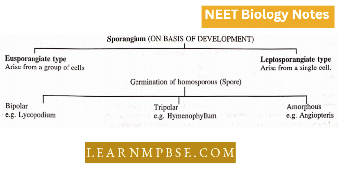
- Eusporangium. Sporangia spore-producing structures develop from a group of initial cells Example in Psilatum Lycopodium, Equisetum.
- Leptosporangium. Sporangia originate from a single superficial cell Example in Marsilea, Pteris In some forms eg Polypodium aureum and Drynaria regidula the leaves develop prothalli under dim light but sporophytic buds under strong light.
- Rhynia was discovered from Rhyncihert Valley in
Scotland by Mackie (1913). - Each leaf of Selaginella bears on its upper surface near its base a small membranous, tongue-like outgrowth ligule.
- Large greatly vacuolated cells for water absorption are called glossopodium.