NEET Biology Plant Growth and Development Basic Features Of Development
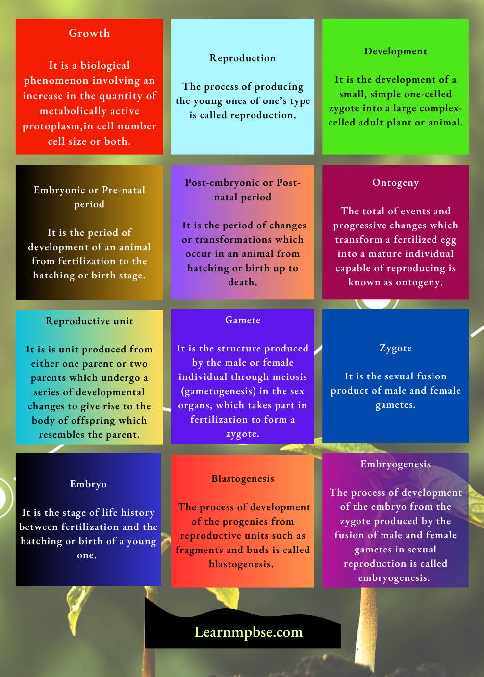
- Growth: It is a biological phenomenon involving an increase in the quantity of metabolically active protoplasm, accompanied by an increase in cell number cell size or both.
- Reproduction: The process of producing the young ones of one’s type is called reproduction.
- Development: It is the development of a small, simple one-celled zygote into a large complex-celled adult plant or animal.
- Embryonic or Pre-natal period: It is the period of development of an animal from fertilization to the hatching or birth stage.
- Post-embryonic or Post-natal period: It is the period of changes or transformations which occur in an animal from hatching or birth up to death.
- Ontogeny: The total of events and progressive changes which transform a fertilized egg into a mature individual capable of reproducing is known as ontogeny.
- Reproductive unit: It is is unit produced from either one parent or two parents which undergo a series of developmental changes to give rise to the body of offspring which resembles the parent.
- Gamete: It is the structure produced by the male or female individual through meiosis (gametogenesis) in the sex organs, which takes part in fertilization to form a zygote.
- Zygote: It is the sexual fusion product of male and female gametes.
- Embryo: It is the stage of life history between fertilization and the hatching or birth of a young one. It is derived from the fertilized egg or zygote. The embryo is diploid. It is formed as a result of fertilization and subsequent changes.
- Blastogenesis: The process of development of the progenies from reproductive units such as fragments and buds is called blastogenesis. The process is common in those individuals who are produced by asexual reproduction.
- Embryogenesis: The process of development of the embryo from the zygote produced by the fusion of male and female gametes in sexual reproduction is called embryogenesis. This process occurs in individuals who reproduce by sexual reproduction.
Read and Learn More NEET Biology Notes
- Gametogenesis: The production and differentiation of male and female haploid gametes from gonads.
- Fertilization: The union or syngamy of haploid nuclei of male and female gametes to form a diploid zygote is called fertilization.
- Cleavage and Blastula: A series of repeated mitotic divisions of an egg that form a cluster of balls of cells (blastomeres) is called blastula.
- Gastrulation: The morphogenetic movements of cells of the blastula to give rise to germinal layers, ectoderm, endoderm and mesoderm is called gastrulation. The embryo thus formed with archenteron is called gastrula.
- Growth: Growth is a permanent increase in the synthesis of new nuclear material and cytoplasm.
- Differentiation: It refers to events by which parts become different from one another. It may be morphological, chemical, behavioural, or differentiation.
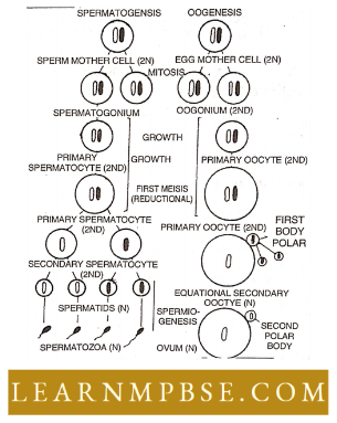
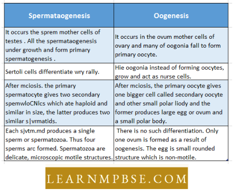
- Spermatogenesis: The process of formation of sperms from the sperm mother cell of the testis. It involves the following phases:
Plant Growth And Development Notes For Neet
Multiplication phase: The sperms are formed from the sperm mother cells, present in the germinal layer of seminiferous tubules of the testis. Some of the mother cells enlarge and divide mitotically to form spermatogonia.
- Growth phase: Some of them enter a period of growth and are called primary spermatocytes which are diploid.
- Maturation phase: These cells divide meiotically to form two haploid, secondary spermatocytes. Each secondary spermatocyte again divides. Thus one primary spermatocyte forms four haploid spermatids.
- Spermiogenesis (Spermateliosis): The process of conversion of spermatid into motile spermatozoa is called spermiogenesis.
- It involves movements of cell organelles characteristics of mature sperm such as shrinking of nucleus forming the head, Golgi complex gathering in front of nucleus forming acrosome, change in centriole as distal centriole from the axial filament, mitochondria form spiral sheath and middle piece.
- All these changes are aimed at keeping the spermatozoa small, light and motile. Oogenesis. The process of formation of the ovum from the ovum mother cells of the ovary. It is similar to spermatogenesis and completed in three phases viz. multiplicative phase, growth phase and maturation phase.
Similarities between Spermatogenesis and Oogenesis:
- Both processes start with primordial germ cells derived from the germinal epithelium of gonads and occur inside the gonad.
- Both the processes are completed in three phases i.e. multiplication, growth and maturation and show meiosis in the maturation phase.
- Both processes produce haploid gametes.
Difference Between Spermatogenesis and Oogenesis:
NEET Biology Plant Growth and Development Structure Of Ovum
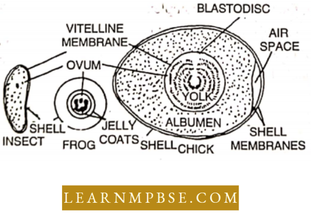
In all the animals, ova arc single-celled. The ovum is surrounded by a primary membrane called vitelline membrane which is produced after the release from the ovary, the secondary and tertiary membranes are secreted by the Graafian follicle or lining of the oviduct. These layers may be albuminous as in amphibians or may be porous, calcareous as in birds.
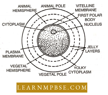
- Most animal eggs are spherical or oval, non-motile but on close examination it is noted that one pole is different from the other. The pole from which polar bodies are given off is called the animal pole while the opposite is termed as vegetal pole.
- Thus it is said to have polarity. Thus various cytoplasmic substances are unequally distributed along the axis. The nucleus, also called a germinal vesicle having a chromatin network is bounded by a nuclear membrane.
- It also contains prominent nucleolus. All animal eggs contain some reserve material to provide food called yolk. The cytoplasm of the ovum is called ooplasm. It lacks a centrosome but contains cortical granules derived from Golgi bodies in its peripheral region termed as cortex.
NEET Biology Plant Growth and Development Functions Of Ovum
- It contributes female’s haploid set of chromosomes.
- After fertilization, forms a zygote which gives rise to young animals by development.
- Egg Membranes Protect The Developing Embryo.
- The eggs are of different sizes. The smallest egg is that of the mouse (0.075 mm) find the largest is that of the ostrich bird (175 mm).
NEET Biology Plant Growth and Development Types Of Eggs
1. Types of eggs based on amount or yolk.
- Alecvithal cups. Yolk absent c.g. Mctathcrian and cutlierian mammals.
- Leithal eggs. Eggs contain yolk. They are of three types.
- Microlcoithal or olipolceithul Eggs: Microlcoithal Eggs contain very small amounts of yolk
- Examples: Hydra, and sea urchin. Amphioxus, Tunicatcs.
- Mesolccithal Eggs: Mesolccithal Eggs contain a moderate amount of yolk
- Examples: Earthworm, Dipnoi fishes, and amphibians.
- Macrolecithal or mepalecithal or polylccithal eggs: Macrolecithal Eggs contain large quantities of yolk
- Examples: birds, fishes, reptiles, prototherian mammals and insects.
- Microlcoithal or olipolceithul Eggs: Microlcoithal Eggs contain very small amounts of yolk
2. According to the distribution of yolk:
Eggs are of the following types
- Home-lethal or Isolecithal eggs have evenly distributed yolk
- Example: Porifers, Amphioxus, Echinodemiates.
- Heterolecithal Or Anisolecithal Eggs: Have Localized Yolk.
- They Are Of The Following Types
- Telolecithal eggs: Telolecithal eggs in amphibians— yolk concentrated towards one side.
- Meiolecithal eggs: Meiolecithal eggs of birds and reptiles are large and yolk occupies the entire ooplasm.
- Centrolecithal eggs: Centrolecithal eggs in insects have yolk in the centre with cytoplasm forming a thin layer around.
- They Are Of The Following Types
3. Types Of Eggs According To Covering:
- Cleidoic eggs. These eggs are fully laden with yolk and surrounded by albumen and water-proof shells made up of calcium
- Example: Eggs of reptiles and birds.
- Non-cleidoic eggs: Non-cleidoic eggs In such a shell is absent.
4. Types Of Eggs According To The Type Of Development:
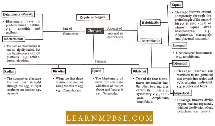
- Determinate or mosaic eggs. In such eggs, cleavage is of a determinate type and each part of the egg has a definite fate
- Examples: Porifers, Platyhelminthes, annelids, arthropods and molluscs.
- Indeterminate or regulative eggs. In this type, each early blastomere on separation from others may give rise to a complete embryo
- Example: echinoderms and chordates.
Neet Biology Plant Growth And Development Pdf
NEET Biology Plant Growth and Development Egg Membranes Or Coats
Eggs possess unique coats beside the plasma membranes. They safeguard the eggs from predators and physical harm while supplying nourishment.
- The primary membrane secreted by the egg is the vitelline membrane.
- The secondary membrane is constituted by follicular cells of the ovary that encase the ovum. Chorion in the egg of Herdmania and zona pellucida in the mammalian egg.
- Tertiary membrane produced by the oviduct’s gland. Jelly-like coating around the eggs of fish and amphibians, together with albumen, shell membranes, and the shell encasing the eggs of reptiles and birds.
NEET Biology Plant Growth and Development Structure Of Mammalian Sperm
Structure. A mammalian sperm is a minute, microscopic, flagellated and motile gamete with no nutritive material, protective envelopes and most cell organelles like ribosome, endoplasmic reticulum, etc. It is formed of four parts, each performing a specific function:
1. Head: The shape of the head varies in different mammals. It is generally oval and flat (in man, bull, rabbit). The head is formed of two parts :
- Acrosome (Gr. Akron = extremity; soma = body): It is a small pointed structure present at the tip of the nucleus. It is formed from a part of the Golgi body of the spermatid. During the sperm entry, the acrosome secretes a lytic enzyme called hyaluronidase which helps in the penetration of the ovum.
- Nucleus: It is generally long, narrow and pointed but is flat and oval in human sperm. It is formed by condensation of nuclear chromatin of spermatid and loss of RNA, nucleolus and acidic proteins. Chemically, the nucleus is formed of deoxyribonucleoprotein (DNA + basic proteins). It is the carrier of genetic information.
The acrosome and anterior half of the nucleus are covered by a fibrillar sheath galea.
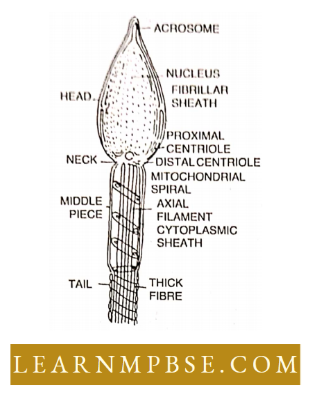
2. Neck: It is the smallest part of spermatozoan and may be indistinct. It is formed of two centrioles perpendicular to each other and is formed from the centrosome of the spermatid.
- Each centriole is a microtubular triplet structure having a 9 + 0 arrangement. The proximal centriole lies in a depression in the posterior surface of the nucleus and is perpendicular to the main axis of the sperm. The distal centriole is along the longitudinal axis of the sperm.
- Centrioles form a spindle for the first cleavage of the zygote. Distal centriole acts as a basal body and gives rise to the axoneme of the sperm tail.
3. Middle piece: It lies behind the neck and is cylindrical in human sperm.
- It is formed of a mitochondrial spiral called nebenkern around the proximal part of the axoneme.
- The middle piece is a powerhouse of sperm.
- The posterior half of the nucleus neck and middle piece of sperm is covered by a sheath called a manchette.
- Tail (Flagellum) It is the longest part of sperm. It is slender and tapering.
It comprises two parts: Central contractile and microtubular, axoneme or axial filament, and outer protoplasmic sheath.
- Axoneme is formed of 11 proteinous microtubules arranged in a 9 + 2 manner.
- Sometimes, a ring centriole may be present at the junction of the middle piece and flagellum.
- The tail shows lashing movements which provide a forward push to the sperm.
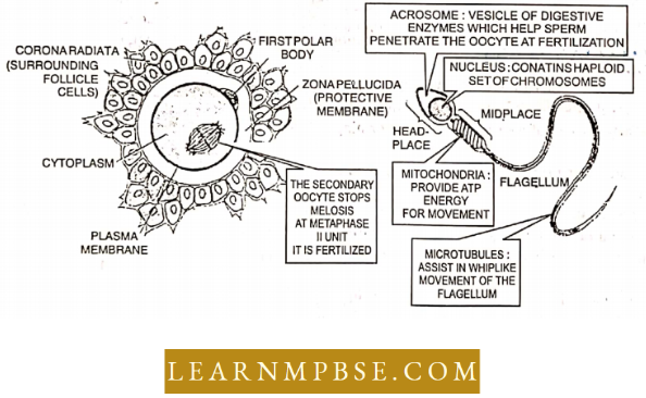
4. Viability: It is the period up to which the sperm can fertilize an ovum. The viability of human sperm is about 24 hours.
NEET Biology Plant Growth and Development Fertilization
Fertilization. It is a fitness by which male and female gametes come close and fuse to form a zygote. The union of two gametes is termed syngamy and the intermixing of nuclei is amphimixis.
Steps Involved in fertilization are :
- Encounter of spermatozoa and ova: It may occur inside the body (internal fertilization) or outside the body(external fertilization).
- Capacitation and contact. Sperm (antifertilizin) and ovum (fertilizin) show fertilizin- antifertilizin reaction which is highly specific for a species.
- Acrosome reaction and penetration: Golgi bodies of acrosome secrete enzymes.
hyaluronidase which dissolves the membranes of the ovum so that materials of sperm can penetrate. - Activation of ovum: Immediately after the apical tip of the acrosomal tubule touches the egg plasma membrane, a fusion of both membranes takes place and a single continuous mosaic membrane is formed.
Thus plasma membrane of both gametes becomes continuous and forms a zygote. At this time certain changes occur in the cytoplasm of the egg which are collectively termed as activation of the ovum. It includes the following events.
- Fertilization cone formation: Coming in contact with the acrosomal filament of spermatozoan, the cytoplasm of the egg bulges forward to produce a conical projection, the fertilization cone. It gradually engulfs the spermatozoan.
- Cortical reactions and fertilization membrane formation: It may differ from one group of animals to the other, but, in most groups, it fundamentally culminates into the formation of a membrane, called fertilization membrane, outside the egg plasma membrane. This membrane blocks the entrance of late-arriving spermatozoa.
- Metabolic activation: Following metabolic changes occur in the egg at the time of fertilization.
- Changes in the plasma membrane: The permeability of the egg plasma membrane increases for the molecules of water, glycol and ions of K+, P04-3 etc. Its electrical potential becomes more positive in the beginning but more negative later on.
- Ionic changes: The concentration of cations such as Na+, Ca++ and K+ changes during fertilization.
- Changes in coenzyme: During fertilization, the inactive enzyme NAD-kinase becomes active and changes NAD into NADP and NADPH by phosphorylation.
- Respiratory changes: Fertilization increases the rate of respiration in those eggs in which maturation is completed before fertilization (e.g., sea urchin) and decreases in those eggs in which fertilization occurs at the first maturation division (e.g., Chaetopterus).The respiratory rate increases for the release of more ATP.
- Change in the rate of protein synthesis: During fertilization, the inhibitor enzymes are removed to initiate an action of protein synthesis.
- Initiation of mitosis: It takes place due to the following agencies :
Immediately after fertilization, the rate of DNA synthesis increases many- folds and so also the uptake of cytoplasmic DNA Polymerase enzyme, which is required in the biosynthesis of DNA. The sperm introduces its centriole and by contributing a second centriole it initiates the formation of a spindle.
Plant Growth Regulators NEET Study Material
- Migration of pronucleus and amphimixis: To perform the act of amphimixis, the sperm nucleus has to perform two activities i.e., it has to become pronuclei and to migrate v from the site of penetration to the site of amphimixis. The fusion of male and female pronuclei is termed amphimixis.
- Significance Of Fertilization:
- It stimulates the egg to complete its maturation.
- The fusion of male and female pronuclei in fertilization restores the diploid number of chromosomes and activation of secondary oocytes results in a mature form of ovum. The ovum which is in a quiescent state with a low metabolic rate restores its normal metabolic activity.
- Fertilization initiates cleavage or segregation.
- The combination of the chromatin material from two different parents forms the physical basis of biparental inheritance and variation.
- The centriole of sperm initiates the first division of zygote timelines and further entry of sperm is checked by the fertility/action membrane.
Ovipary, Vivipnry and Ovovivipnry:
- Osipary: It is the development of an embryo inside its egg delivered outside the body. The fertilization may be external or internal.
- Vivipnry: It is the development of an embryo inside the uterus of a female connected using the placenta. The fertilization is internal. The female gives birth to the young one.
- Ovovivipnry: The phenomenon of retaining eggs and the development of embryos in the body till birth without forming any organic connection for obtaining extra nourishment i.e. no placenta formation. Fertilization is internal.
NEET Biology Plant Growth and DevelopmentCleavage
Cleavage: The series of divisions that occur inside the fertilized egg or zygote to transform it into a multicellular body called blastula is called cleavage. They produce blastomeres.
Characteristics of Cleavage:
- Cleavage divisions are mitotic and occur one after the other. However, the rate depends upon species, amount of yolk and temperature.
- During cleavage, growth does not take place and the size and volume of the embryo remain the same and the size of blastomeres is reduced.
- The nuclear and cytoplasmic ratio becomes very low for the zygote and as a result cleavage increases.
- During cleavage general shape of the embryo does not change.
- All blastomeres divide simultaneously during early cleavage.
- For rapid nuclear division, there is a great increase in the synthesis of DNA.
- O2 consumption increases during cleavage.
The cleavage division differs from ordinary mitotic division in the following ways :
- The cleavage division is not followed by the growth of daughter cells before they divide but during ordinary mitotic division, each division is followed by a growth of daughter cells before the next division starts.
- The rate of cleavage depends in different species on temperature but during mitosis, it is not so.
- During cleavage nuclear and cytoplasmic ratio is changed.
- During cleavage, the blastomeres divide simultaneously but in ordinary mitosis all the daughter cells divide approximately at the same time.
- The cleavage differs from ordinary mitosis of large-sized zygotes, and it is meant basically to reduce the size of blastomeres.
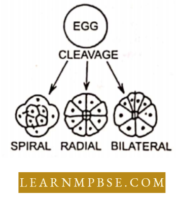
Pattern of Cleavage: Three common patterns of cleavage are radial, bilateral and spiral cleavage.
- Radial cleavage: In radial cleavage, the successive cleavage planes cutting straight through the egg are at a right angle to one another. The resultant blastomeres become symmetrically, exposed around the animal-vegetal axis. Such type of cleavage occurs in sponges, coelenterates and a few molluscs.
- Bilateral cleavage: In this pattern of cleavage, the blastomeres are so arranged that the right and left sides become apparent, e.g., Cephalopods, a few Echinoderms and Vertebrates.
- Spiral cleavage: In this pattern of cleavage, the furrows are so formed that the blastomeres are arranged in a spiral manner around the animal-vegetal axis e.g., Flatworms, annelids and molluscs.
Planes Of Cleavage During cleavage, different cleavage furrows may divide the egg into different planes.
- Meridional plane. Cleavage furrow passes through the centre of the animal-vegetal axis and bisects both the poles of the egg. e.g., Frog.
- Vertical plane. Cleavage furrow passes in a direction from the animal pole towards the vegetal pole. For. first cleavage furrows of Amin calva and chick.
- Equatorial plane. It bisects the egg at right angles to the main axis and halfway between the animal and vegetal poles, e.g…… first cleavage plane of eggs of higher mammals.
- Latitudinal plane. It is similar to equatorial, but it courses through the cytoplasm on either side of the equatorial plane, e.g., the third cleavage planes of Amphioxus and frog.
Neet Biology Plant Growth And Development Pdf
Types of cleavage: The amount and pattern of distribution of yolk determine the type of cleavage. It is of the following types :
- Holoblastic cleavage: In alecithal, isolecithal and slightly telolecithal eggs, the zygote and the blastomeres divide completely. It results in a cocloblastula i.e., a blastula with a central cavity called the blastocoel. Holoblastic cleavage is of two types
- Equal holoblastic cleavage: The cleavage produces approximately equal-sized blastomeres throughout. It occurs in alecithal eggs of rabbits and microlecithal eggs of sea squirts, amphioxus, marsupials and other placental mammals.
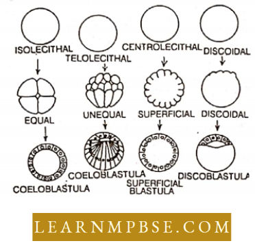
- Unequal holoblastic cleavage: This cleavage in later divisions gives rise to unequal blastomeres, small micromeres and large megameres. It occurs in telolecithal eggs of bony fishes and amphibians.
- Meroblastic Cleavage: In this type of cleavage, division occurs only in the small amount of metabolically active cytoplasm. The yolk remains undivided. It is found in macrolecithal eggs. It is of two types :
- Discoidal Cleavage: It is confined to the active cytoplasm located at the animal pole and occurs in isolecithal eggs of reptiles, birds and egg-laying mammals. It results in a blastula called a disco blastula.
- Superficial Cleavage: It occurs in the centrolecithal eggs of insects. The nucleus placed in the centre of the yolk divides repeatedly to form many daughter nuclei which migrate to the peripheral yolk-free cytoplasm. It then cleaves into many uninucleate blastomeres arranged around the yolk. It results in a superficial blastula consisting of single-layered epithelium enclosing yolk instead of blastocoel.
Significance of cleavage:
- It produces a large number of cells from a single-celled zygote by repeated divisions and results in the formation of the embryo. , .•
- It prepares the embryo for the migration of cells to their fixed position during gastrulation.
- Preparation and initiation of the process of cell differentiation.
NEET Biology Plant Growth and Development Gastrulation
Gastrulation. It is a phase of embryonic development during which cell movements establish the three primary germinal layers and initiate morphogenesis.
The most important features of gastrulation in all animals are :
- Complex but orderly and irreversible morphogenetic movements involving large masses.
- The rhythm of cellular division is slowed down.
- Growth if any is insignificant.
- The nuclei become more active.
- Formation of archenteron surrounded by endoderm.
- The type of metabolism changes and the rate of oxidation is intensified.
- Chemodifferentiation starts with the synthesis of new proteins.
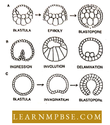
- Epiboly: It involves stretching and spreading movements of ectoderm-forming cells to surround the embryo except at one pole called the blastopore.
- Emboly: It is the shifting or migration of the cells in such a way as to form internal layers of the mesoderm and endoderm of the embryo. It can occur by five methods.
- Invagination: It is the simplest type of emboly. In this method, the vegetal pole side of the blastula simply caves in or pushes into the interior e.g. Amphioxus and Invertebrates.
- Involution: It is the active rolling in of the endodermal and mesodermal cells from the surface to the interior through blastopore e.g., Frog.
- Ingression: It is the proliferation of cells from the inner surface of the blastoderm into blastocoel which gets filled up with new cells and results in the formation of solid gastrula. The archenteron appears later on by splitting up of internal mass of cells. It may be unipolar as in Obelia or apolar as in Hydra.
- Delamination: Separation of sheets of cells to form two layers called epiblast and hypoblast e.g. chick, mammals.
- Convergence: It involves the migration of blastomeres from the outer surface towards the blastoporal lips.
NEET Biology Plant Growth and Development Fate Of There Germinallayers
Germinal layers:
Ectoderm, Mesoderm and Endoderm are the three primary germinal layers of the embryo as all the organs differentiate and develop from these layers.
NEET Biology Plant Growth and Development Morphogenesis And Nieekrentlation
Every living organism starts its life from a one-celled structure, a zygote, formed by a union of male and female gametes. From this one-celled stage, a multicellular stage is established by groups of processes of which morphogenesis and differentiation are outstanding.
Morphogenesis: It involves the formation of various structures in an individual organism resulting in the establishment of a young one.
There are two main processes of morphogenesis, namely growth and form:
- Growth: Molecular growth increases the size of the individual cell. The mitotic division of cells, the most significant process of growth involves the increase in the number of cells and eventually the increase in bulk and complexity of the organism.
- Form: The growth leads to the form of an individual plant or animal. The resultant of growth establishes various structures and organs. When a structure or an organ has reached its maximum limitation of size, further growth stops. The form is determined by polarity and symmetry. Polarity is of the three dimensions. In an organism one exerts dominance.
The head exerts dominance over the rest of the body. Due to polarity, there is a differentiation of anterior, posterior, right and left sides. Symmetry is the regularity of the arrangement of parts in an organism.
Differentiation: This involves the operational and functional components of development. The zygote divides repeatedly finally establishing millions of cells all having the same types of genes. Yet these cells differentiate into various organs. A group of cells at an early stage maintain certain functions while in the later part of life take up different functions.
Up to a stage the cells are capable of giving rise to any structure i.e. totipotent and after a time when a cell produces special types of cells, there cannot be any further transformation of the cell. The final stage reaches specialisation so that the cell is set apart to perform a definite function.
Plant Hormones And Growth Regulators Neet Notes
Cell differentiation may come about in three methods :
- The developing differences among cells might be the result of progressive changes in gene action. Further, gene activity patterns might change differently in different cells resulting in differentiation.
- While the gene action might remain constant, the cytoplasmic operations may become progressively altered.
- Nuclear and cytoplasmic changes might occur reciprocally.
- The differentiation may start from the molecular level leading to cellular differentiation.
NEET Biology Plant Growth and Development Formation Of Three Germinal Layers In Mammal
Development of three germinal layers. In viviparous mammals, fertilization occurs internally.
The subsequent processes transpire during the creation of the three germinal layers:
1. Cleavage:
The zygote undergoes holoblastic cleavage in the fallopian canal.
2. Morula:
A compact sphere of cells is generated through successive cleavage divisions.
3. Blastula:
(Blastodermic vesicle or Blastocyst) A chamber known as the blastocoel emerges in the developing embryo. The blastodermic vesicle is encased by an external cellular layer known as the trophoectoderm. The blastodermic vesicle exhibits an interior aggregation of cells resulting from differentiation. This aggregation of cells is referred to as the inner cell mass.
4. Gastrulation:
At this stage, the morphogenetic movements of the cells, such as epiboly and emboly, occur in small aggregates or sheets.
Consequently, three germinal layers are established:
- Endoderm
- Mesoderm
- Ectoderm
1. Development of Endoderm:
Certain cells from the inner cell mass undergo delamination.
- Cells migrate in layers or aggregates within the blastocoel. These represent the prospective endodermal cells.
- These cells organize themselves as the second layer, situated between the inner and outer layers of the blastodermic vesicle.
- The blastocoel vanishes, and a new hollow emerges. The newly formed cavity is referred to as the archenteron.
- The residual mass of embryonal knob cells becomes convoluted and systematically organized to create an embryonic disc.
2. Formation or mesoderm:
The cells at the caudal end of the embryonic disc initiate proliferation and subsequently detach from the disc to produce mesoderm.
3. Development of ectoderm:
Following the detachment of mesodermal cells from the embryonic disc, the remaining cells in the disc organize into a separate layer, forming the ectoderm.
NEET Biology Plant Growth and Development Placenta
- Placenta: A vascular organ that unites the foetus to the wall of the uterus in all mammals except marsupials and monotremes is called the placenta and its formation is plantation.
- Structure or Kpithelio-shoring placenta: Epitheliochorial is the simple and basic type of placenta formed in marsupials, pigs, horses, etc. In this case, some of the components are derived from the uterine wall of the mother while the other parts are derived from the foetus itself.
- Maternal components are (a) mucous membranes of the uterine wall (b) uterine connective tissue (maternal mesenchyme) (c) endothelium of maternal blood capillaries.
- Foetal components are A wall of foetal blood capillaries and foetal connective tissue. Foetal chorionic epithelium.
- Functions of the placenta: The placenta serves primarily as an organ that permits the interchange of material earned in the blood of the mother and foetus. The main functions are :
- Nutrition: Supply of nutrient materials to the foetus. Respiration. Supply of 02 to the foetus and receive C02 back from it. Excretion. Fluid nitrogenous waste products escape through the placenta. Barrier. The placenta is a barrier-like semipermeable membrane.
- Storage: The placenta stores fat, glycogen and iron for the embryo before die formation of the liver. Hormonal function. The placenta secretes extra-ovarian hormones estrogen and progesterone in females during pregnancy that serve to maintain the foetus.
Classification of the placenta: The placenta is of five types depending upon the number of tissue layers that separate the foetal blood from maternal blood.
Epitheliochorial placenta (all six layers) e.g. horse, pig and donkeySyndesmochorial placenta (five layers present, uterine epithelium eroded e.g., cow, goat, sheep Endotheliochorial placenta (four layers present-uterine epithelium and connective tissue eroded) e.g. dog and catHaemochorial placenta (three layers present-material three layers eroded) e.g, primates including human andHaemoendothelial placenta (all the maternal and two foetal tissue layers eroded) e.g. rodents and rabbits.
Previous Year Neet Questions On Plant Growth
Types of placentae according to the distribution of villi on chorion.
- Diffuse placenta: Chorionic villi remain scattered all over the surface of the chorion e.g., pig, horse, lemur.
- Cotyledonary placenta: The villi are found in groups or patches e.g. cattle, sheep, deer
- Zonary placenta: The villi are developed in the form of a belt or girdle e.g. cats, dogs Discoidal placenta. The villi continue developing only on one side e.g. mouse, rat, human, monkey.
- The placenta is of three types, viz. Allantoic, yolk sac and chorionic placenta: about the foetal membrane that takes part in its formation.
NEET Biology Plant Growth and Development Vitellogenesis
The synthesis of yolk is termed vitellogenesis. Recent investigations have revealed that yolk is not necessarily synthesized in oocytes but produced elsewhere in some extra ovarian tissue such as fat bodies in insects.
- In fishes, amphibians and also in gastropod molluscs, its synthesis takes place inside modified mitochondria.
- In other vertebrates, yolk is produced in liver cells. It is transported to the ovarian follicle by blood in the soluble form. From these cells of the follicle, the oocyte takes the yolk by pinocytosis.
- The Golgi complex and endoplasmic reticulum of oocytes transfer these yolk components to modified mitochondria and convert them into insoluble forms.
- Composition of yolk: The yolk is the reserve food material in the egg. It is composed of proteins, phospholipids and neutral fats. The yolk may be a protein yolk if it contains mainly protein. If the yolk consists mainly of phospholipids, neutral fats and small fats, it is termed a fatty yolk. Both kinds of yolk are present side by side in the eggs but protein yolk is the main constituent of many invertebrates and chordates.
- Utilization of yolk: In the isolecithal eggs Example: frog) the yolk gets distributed to all the daughter cells at the time of cleavage. However, at later stages, some cells may have more yolk due to unequal division. The yolk is broken down into simpler substances and utilized in the synthesis of new protoplasmic constituents.
In the acrolectal egg of birds, the yolk is absorbed by diffusion in the early stages. Later on, a yolk sac develops around the yolk mass from mesoderm and endoderm. Blood vessels called vitelline arteries and veins connect the yolk sac with the heart. The enzymes from the yolk sac digest the yolk into soluble form, which is then taken to the heart and vitelline veins and then distributed to all parts of the developing embryo.
NEET Biology Plant Growth and Development Hereditary Defects
- Teratology: It is the branch of developmental biology which deals with the study of abnormal development during embryogenesis. The ways and means by which abnormal developments occur during embryogenesis are called teratogenesis.
- List of hereditary defects, Colour blindness harelip (Cleft plate Clubfoot Hole in the heartAbsence of hands, feet, arms or legs (Meromelia) Right half body normal and left like dwarf (Achondroplasia) Mongolism Phenylketonuria Albinism Alkaptonuria Haemophilia Polydactyly Cat cry syndrome (at) Turner’s syndrome and Klinefelter syndrome.
- Identical twins: The twins are of the same sex and are identical in all respects including appearance, size and behaviour.
The identical twins are monozygotic because they develop as a result of fertilization of the single ovum by a single sperm but the zygote divides into two halves and each half gives rise to a child. Each gets attached to the same placenta. - Fraternal twins: The fraternal twins are dizygotic as they are produced due to the fertilization of two different eggs. They may or may not belong to the same sex. They may or may not resemble each other.
- Normally the ovaries release a single egg at a time or if two eggs are released both may be fertilized and then may develop into two separate individuals, thus fraternal twins develop from two different zygotes, hence termed dizygotic twins. They are connected to the uterine wall by a separate placenta.
- Conjoined twins: Twins are generally normal babies, but some twins may be abnormal in shape, structure or development. Some twins may be fused wholly or in part, such fused twins are called conjoined twins. The degree of union may be slight or extensive. Union is by heads and upper trunk.
Previous Year Neet Questions On Plant Growth
NEET Biology Plant Growth and Development Important Notes
- Spermiogenesis: The process of conversion of a spermatid into a functional spermatozoan by the process of differentiation of specialization is called spermiogenesis or spermioteliosis.
- Sertoli cells: These are indifferent cells situated in the lining of seminiferous tubules which provide nourishment to the differentiating spermatozoa. Sperms’s head is kept embedded in Sertoli cells for this purpose.
- External fertilization: The fusion of male and female gametes takes place outside the body of a female animal.
- Internal fertilization: The fusion of male and female gametes takes place inside the body of a female animal.
- Amnion: A thin extra-embryonic membrane forms a closed sac around the embryo in birds, reptiles and mammals.
- Amniota: A collective term for the reptiles, birds and mammals, all of which have an amnion.
- Anamniote: Any animal of which the amnion is absent during embryonic development.
- Amniotic fluid: A substance that fills the amnion to protect the embryo from shock and industry.
- Amniocentesis: A procedure during pregnancy by which the abdominal wall and foetal membrane are punctured with a cannula to withdraw amniotic fluid.
- Amphimixis: The union of nuclei of egg and sperm in sexual reproduction.
- Allantois: A fluid-filled, sac-like extraembryonic membrane lying between the chorion and amnion of reptiles, birds and mammals.
- Allantochorion: The extraembryonic membrane is formed by the fusion of the outer wall of allantois and with the primitive chorion.
- Chemo-difTerentiation: It is the chemical differentiation of cells of the embryo during development.
- Cyto-differences: It is the differentiation of cells ol ciuluyo dining development.
- no-dinViviilintlon: It location of the tissue mill also ballots ns histogenesis.
- Orgnn-dinVivntlntlon: It is tot million of guns limit the embryo.
- Kmhnoule Murillo: The supply of required materials which are essential for the synthesis of unit ami and other compounds ami for the production of energy during the development of the embryo is called embryonic nutrition. The materials required are amino acids, minerals and oxygen.
- Diapause: The sexual rest period in insects is called diapause.
- Kmlometrium: Glandular mucous membrane lining of the uterus.
- Genome: The set of all different chromosomes found in each nucleus of a given species. A haploid nucleus has one genome.
- Germinal vesicle: Nucleus of the animal oocyte during the period of cytoplasmic growth.
- Gestation period: Length of time from conception to birth in viviparous animals.
- lactation: Production of milk.
- Monoccious:fUiscxual or hermaphrodite).
- Mullerian Duct: Oviduct of female gnathostome vertebrate.
- Neoteny: Persistence of the form of a larva or any other stage (larva becomes sexually matured.)
- Example: Axolotl Larva of Salamander in Amphibians
- Nidicolous birds: Those which hatch undeveloped.
- Nymph: Young states of exopterygote insect.
- Oxytocin: A hormone secreted by the posterior lobe of the pituitary gland which causes labour pains (contraction of uterine muscles.).
- Secondary sexual characters: A characteristic of animals which differ between the two sexes.
- Seminal vesicles: Organs which store sperm in males (cf).
- Spermatheca. Organs which store sperm in female ($).
- Syngamy:Union of gametes is termed syngamy.
- Testosterone: A principal male hormone.
- Urethra: Duct leading from the urinary bladder of mammals to the exterior.
- Uterus masculinus: A sac-like structure dorsal to the bladder in male mammals.
- Vitelline membrane: Primary egg membrane secreted by the egg.
- Yolk plugs: A mass of macromeres which closes the blastopore in the developing embryo
- Colostrum. The first milk which comes out from the mother’s mammary glands just after childbirth is known as colostrum. It is rich in calories and proteins. It contains antibodies.
- On average human ejaculate of 3 – 4 ml of semen contains 80-100 million spermatozoa.
- Virus infection of the mother e.g. by the rubella (German measles) virus or exposure to certain chemicals may cause malformation in the developing embryo. Such agents are called teratogens. All the systems in a developing foetus are formed by the three months of pregnancy.
- cv During further development after three months, there was growth and a few minor structural modifications. Extra-embryonic membranes include the yolk sac, amnion, chorion and allantois.
- Yolk sac is also found in like certain fishes Example: Scoliodon), bony fishes and a few amphibians
- Example: Necturus having an egg.
- Amnion, chorion and allantois are formed only in am- niote embryos as their development occurs on land either inside the egg or in the uterus of the mother.
- These are not formed in amniotes as their development occurs in water so there is no problem of desiccation, supply of oxygen and removal of wastes. c& Amnion is an innermost embryonic membrane and never participates in placenta formation.
- c& Chorion is an outermost embryonic membrane and always participates in placenta formation.
- Polarity. The existence of a definite axis in the egg and the embryo is called polarity. In the egg, polarity is indicated by the position of the nucleus and the yolk.
- Inducers (Organizers). The cells that induce or control die developmental fate of the neighbouring cells in an embryo are called inducers or organizers.
c& Inductors. These are the chemicals which the inducers release to guide the fate of the adjacent cells.
NEET Biology Plant Growth and Development Quanta To memory
- Identical twins: Twins are the term used to describe the delivery of two or more infants in a single birth. These may be identical twins, monozygotic twins, fraternal, dizygotic, or non-identical twins.
- Siamese siblings: Conjoined twins are joined at the pelvis, chest, kick, and face. These are monozygotic and have been surgically separated for the first time in Siam.
- In the process of frog oogenesis, a single oogonium generates a single ovum and only two polar bodies.
- In mammals, a fertilization membrane is not formed; however, die enzymes that are released following the formation of the zona pellucid neutralize sperm receptors, preventing any additional sperm from binding to them.
- Transformation: Metamorphosis is the term used to describe the process by which a juvenile individual undergoes a transformation into a morphologically and physiologically distinct adult. There are two varieties of it.
- Metamorphosis that is retrogressive: The transformation of an advanced larva into a degenerate adult, such as Merdmania. Sacculina.
- Metamorphosis in progress: When a simplified larva transitions into an advanced adult, such as a frog.
- Martinis are complimentary: A male and a female heifer that were sexually underdeveloped were united.
- Fate diagram: Diagram illustrating presumptive or potential regions on the blastula’s surface.
- This is accomplished through the utilization of specific essential stains, including neutral red, Nile blue sulphate, and Bismarck brown. It was initially prepared by W. Vogt in 1929.
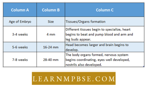
Stages Of Embryonic Development in Human